

Si deseas distinguir tus productos, servicios o ambos de los de otra empresa, es posible que necesites una marca o nombre comercial. Descubre qué son, en qué consiste su procedimiento de registro y qué implica.

Información sobre los plazos de presentación de solicitudes de transformación de marcas de la Unión Europea en marca nacional española. Más información


Si tienes un nuevo dispositivo, producto o procedimiento que resuelva un problema técnico o tenga una ventaja práctica, existen distintas formas de protegerlo en España y en otros países. Descubre cómo hacerlo.

¿Tu innovación reside en la estética, la ornamentación o la apariencia de tu producto? Protégela mediante un diseño industrial. Descubre qué derechos confiere el registro y cómo realizar la tramitación.

Las indicaciones geográficas protegen el nombre de un producto originario de una zona geográfica, a la cual le debe una determinada calidad, reputación u otra característica. Descubre qué son, en qué consiste su procedimiento de registro y qué beneficios conceden.

Las patentes publicadas en todo el mundo son una valiosa fuente de información científica, técnica y comercial.

Si eres emprendedor/a o una empresa y quieres potenciar y mejorar la rentabilidad de tu negocio protegiendo de forma adecuada los activos intangibles de tu organización, en este espacio encontrarás lo necesario.


 169
resultados
169
resultados
 Última actualización
01/04/2026 [08:17:00]
Última actualización
01/04/2026 [08:17:00]


 Resultados 25 a 50 de 169

Resumen de: AU2025260012A1
The present invention relates to a 3D scaffold implant system for mandibular reconstruction, featuring a scaffold structure that includes at least four scaffold segments strategically positioned within the implant, the segments ensuring precise placement within a subject's jawbone and facilitate optimal integration with a surrounding bone tissue; a fluid delivery tube system integrated into each of the scaffold segments to administer a growth factor(s) and an antibiotic directly into the scaffold implant; a pump that facilitates controlled delivery of the growth factors into the scaffold segments; at least three fixation that provide stability to the implant and micro-motors that enable precise alignment of the scaffold implant to conform seamlessly to the natural curvature of the jawbone.

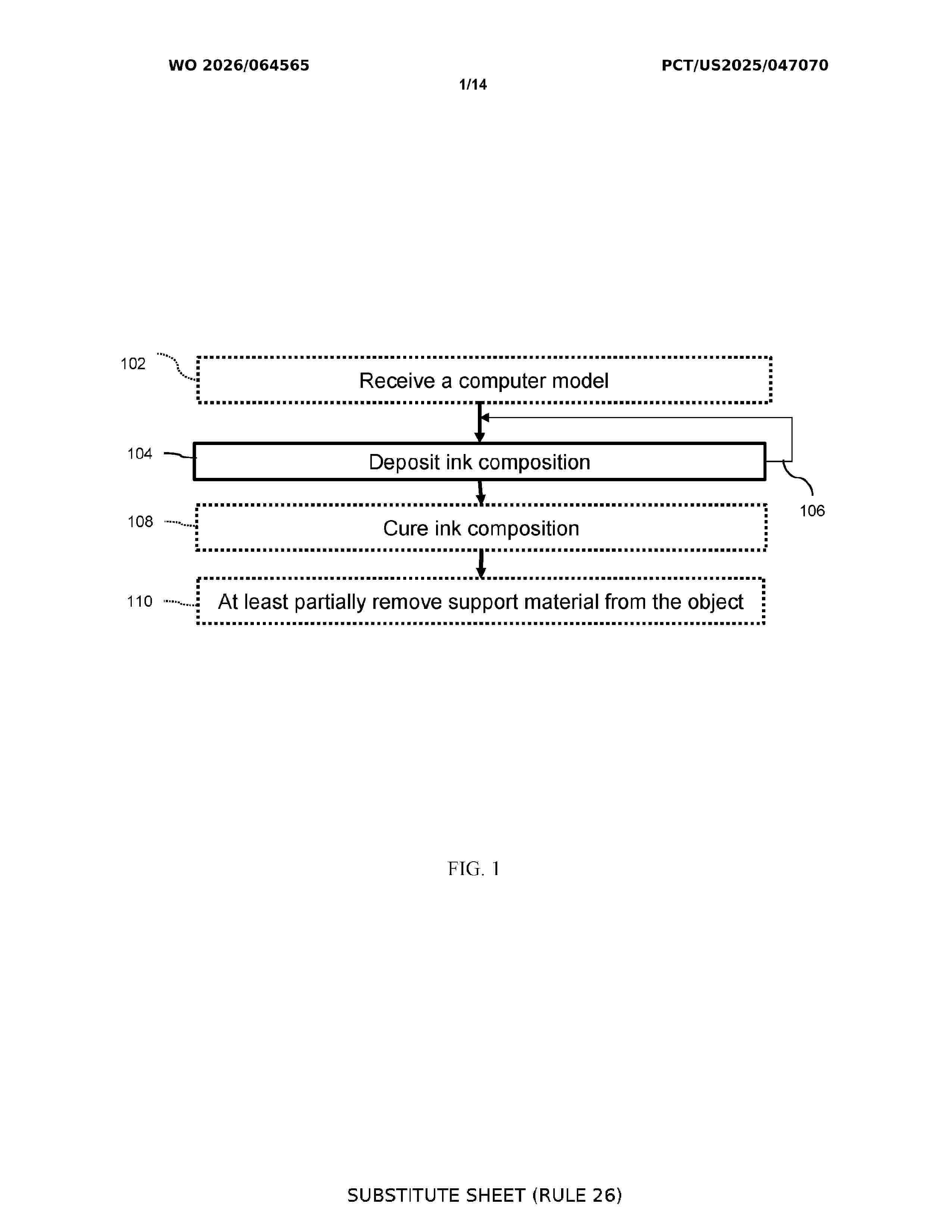
Resumen de: WO2026064565A1
Methods for additive manufacturing, biological structures, cultivated meats, and additive manufacturing systems are provided. The method comprises dispensing an ink composition from a nozzle of an additive manufacturing system. The ink composition comprise adipocytes and a hydrogel. The deposition occurs at a suitable shear stress such that the ink composition can flow through the needle and an integrity of the adipocytes is substantially maintained. The method comprises repeating, as necessary, repositioning of the nozzle and dispensing ink composition from the first nozzle, thereby forming the structure.
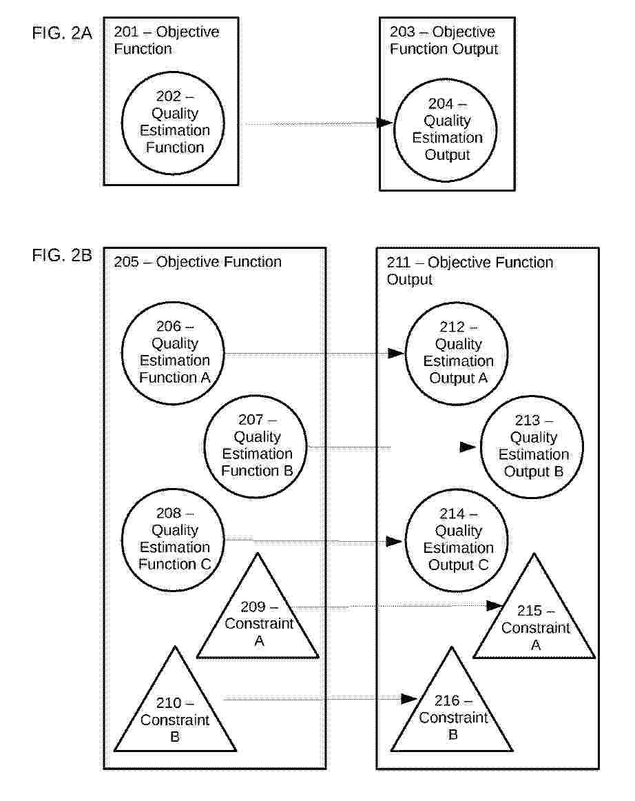
Resumen de: EP4715752A2
Disclosed is a computer-implemented method of generating a dental model based on an objective function output, comprising creating an objective function comprising at least one quality estimation function which trains at least one machine learning method that generates quality estimation output, and an objective function output is the output of the objective function providing a model as an input data to the objective function and generating model-related objective function output; and modifying the model based on the model-related objective function output to transform the model to a generated model, wherein the generated model is the dental model.
Resumen de: JP2023050476A
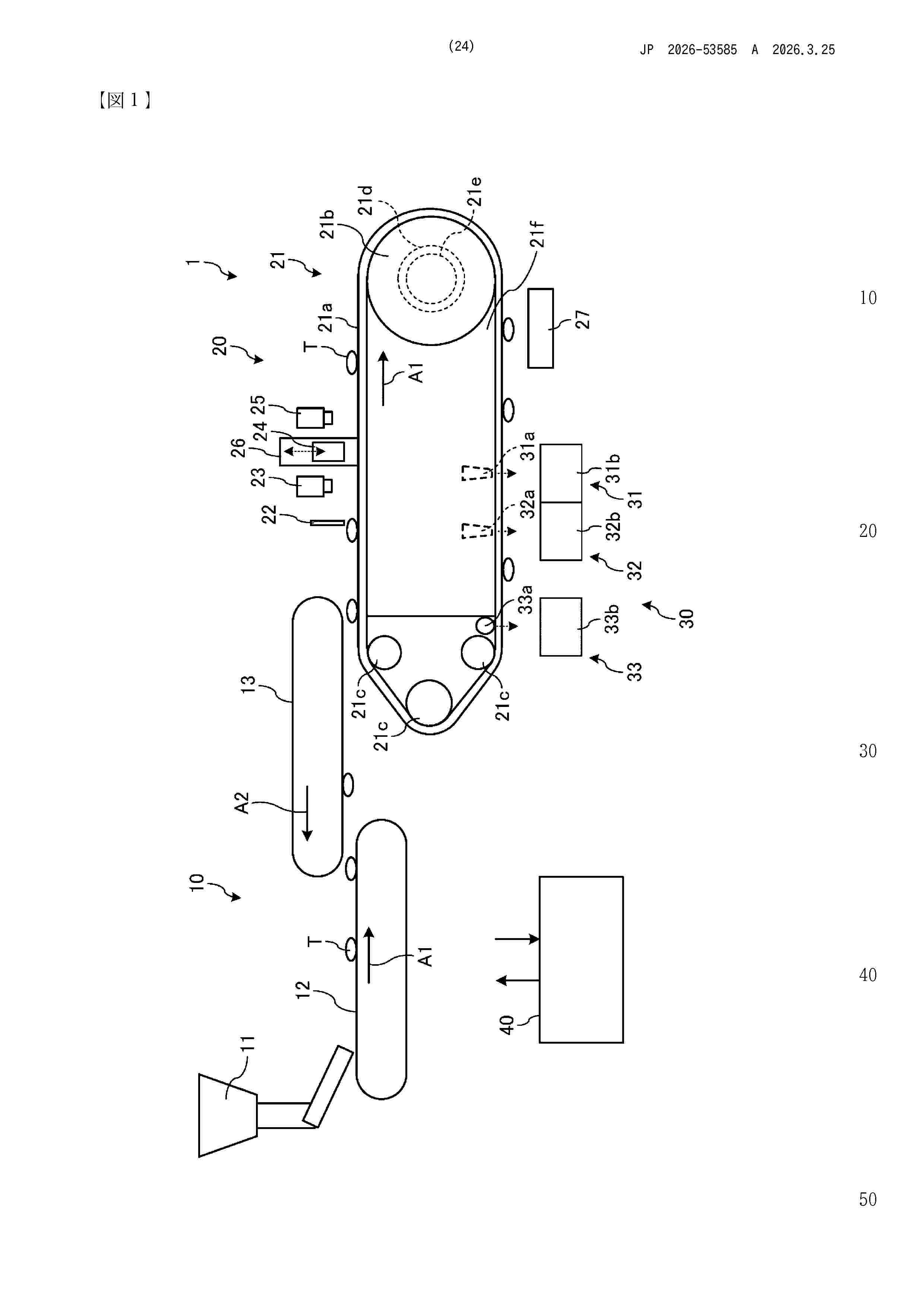
To execute efficient tablet printing.SOLUTION: A tablet printing device 1 includes: a conveyance part 21 for conveying tablets T by a conveyance belt 21a; a detection part 22 for detecting the conveyed tablets T; an inkjet head 24 for discharging ink to the tablets T detected by the detection part 22; a moving mechanism 26 for moving the conveyance belt 21a or the inkjet head 24 so as to change an interval between a surface on a conveyance belt 21a side of the inkjet head 24 and a surface on an inkjet head 24 side of the conveyance belt 21a; and a control part (e.g., a control device 40) for controlling the conveyance part 21 and the moving mechanism 26. The detection part 22 detects height information on the tablets T on the conveyance belt 21a. The control part controls the conveyance part 21 so as to decrease a conveyance speed of the conveyance belt 21a when the height of the tablets T exceeds a predetermined threshold on the basis of the height information, and controls the moving mechanism 26 so as to widen the interval.SELECTED DRAWING: Figure 1
Resumen de: US2022184276A1
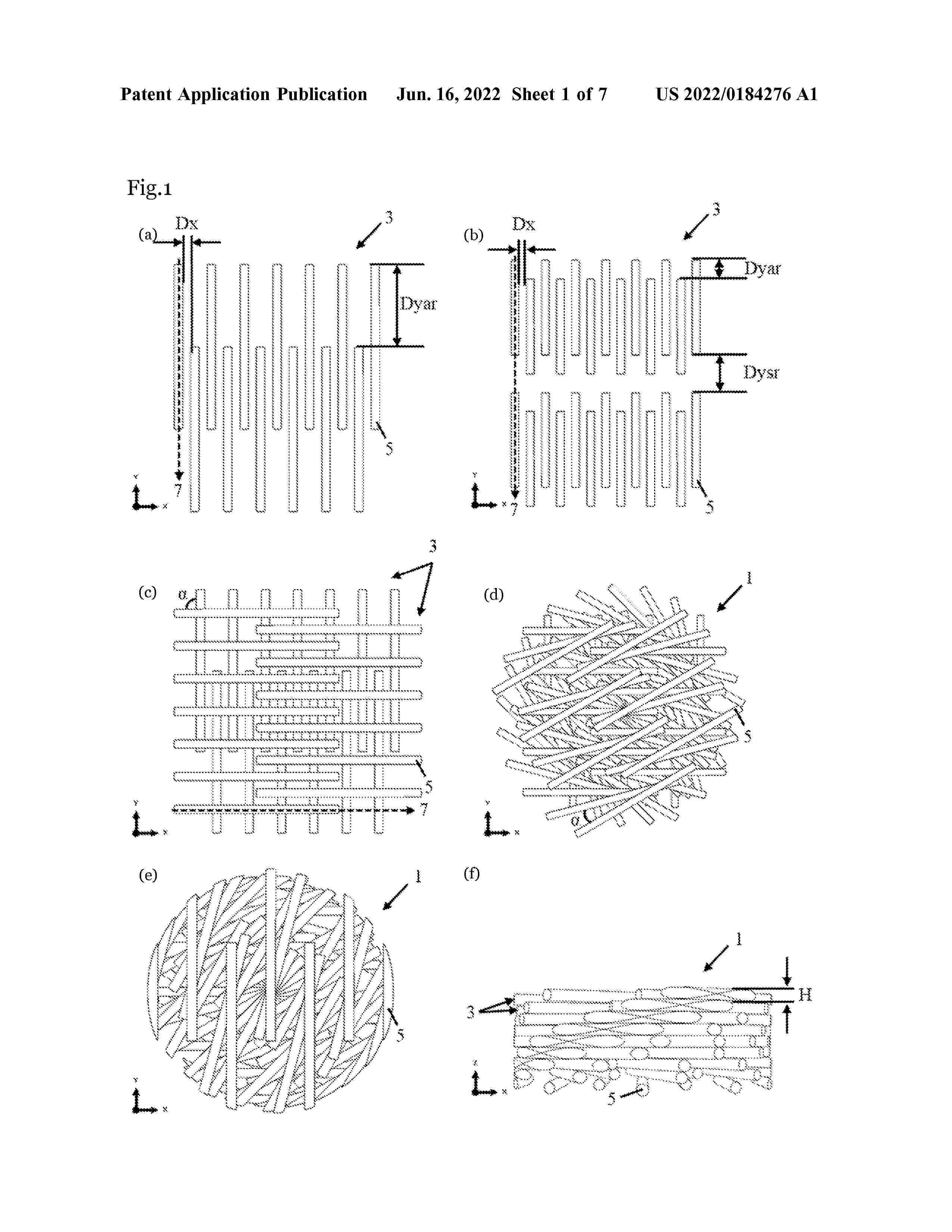
The present disclosure is directed to a degradable 3D-printable scaffold for use in tissue engineering, which scaffold has a combined gradient and staggered structure. Further provided is a medical device for use in tissue engineering, comprising such a scaffold. The present disclosure also provides a method for preparing a scaffold by additive manufacturing, e.g. 3D-printing, a method for in vivo tissue engineering, use of the scaffold in an in vitro cell culture system, in an in vitro method for culturing of cells and/or in an in vitro method for regenerating tissue. Also provided is a scaffold and a medical device for use in a method for in vivo tissue engineering. Further disclosed is a novel degradable copolymer of ε-caprolactone and p-dioxanone, which can be printed without degradation and which is particularly suitable for use as scaffold material in the scaffold and method according to the present disclosure.
Resumen de: GB2700860A
A bioprinting apparatus 100 for 3D printing a model comprises a printing nozzle 115 for dispensing a magnetic or magnetisable bioink for forming the model; a bath 130 containing a model-supporting fluid; a mechanism 110 for moving the nozzle relative to the bath; and at least one magnet 140/145, i.e. a single magnet, a magnet array, magnetisable material or an electromagnet, at or adjacent to the bath 130 suitable for providing a magnetic field 10 in the bath of sufficient strength to influence bioink in the bath and hold bioink in the bath, for example in a stationary manner, until crosslinking occurs. The magnet element may be a magnet array arranged as a Halbach array. Preferably, the magnetic field may hold the bioink at a desired height at which embedded printing is to take place and a controller is operable to control the magnet to move to a different height corresponding to a different layer. The printed material may contain cells, such as eukaryotic cells, and the fluid may contain nutriments for the cells. Printing error correction is described also. Figure 1
Resumen de: CN121240894A
The present disclosure relates to a hydrogel ink comprising a lipid particle, a polynucleotide supported within the lipid particle, and a bio-ink. In one embodiment, the lipid particles comprise extracellular vesicles. The disclosure also relates to hydrogels thereof and their use in treating neurological diseases or conditions or regenerating soft tissue.
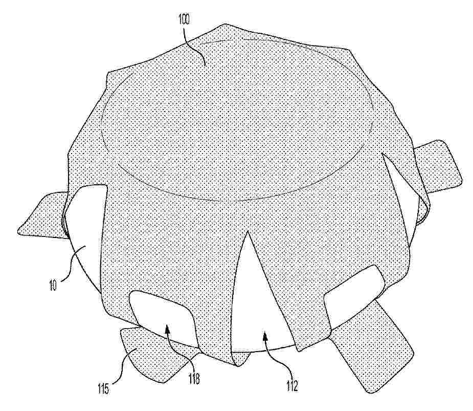
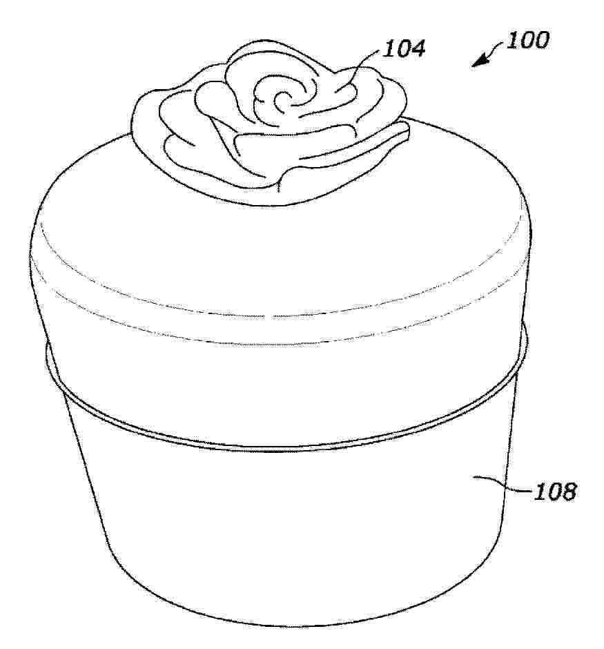
Resumen de: CN121398761A
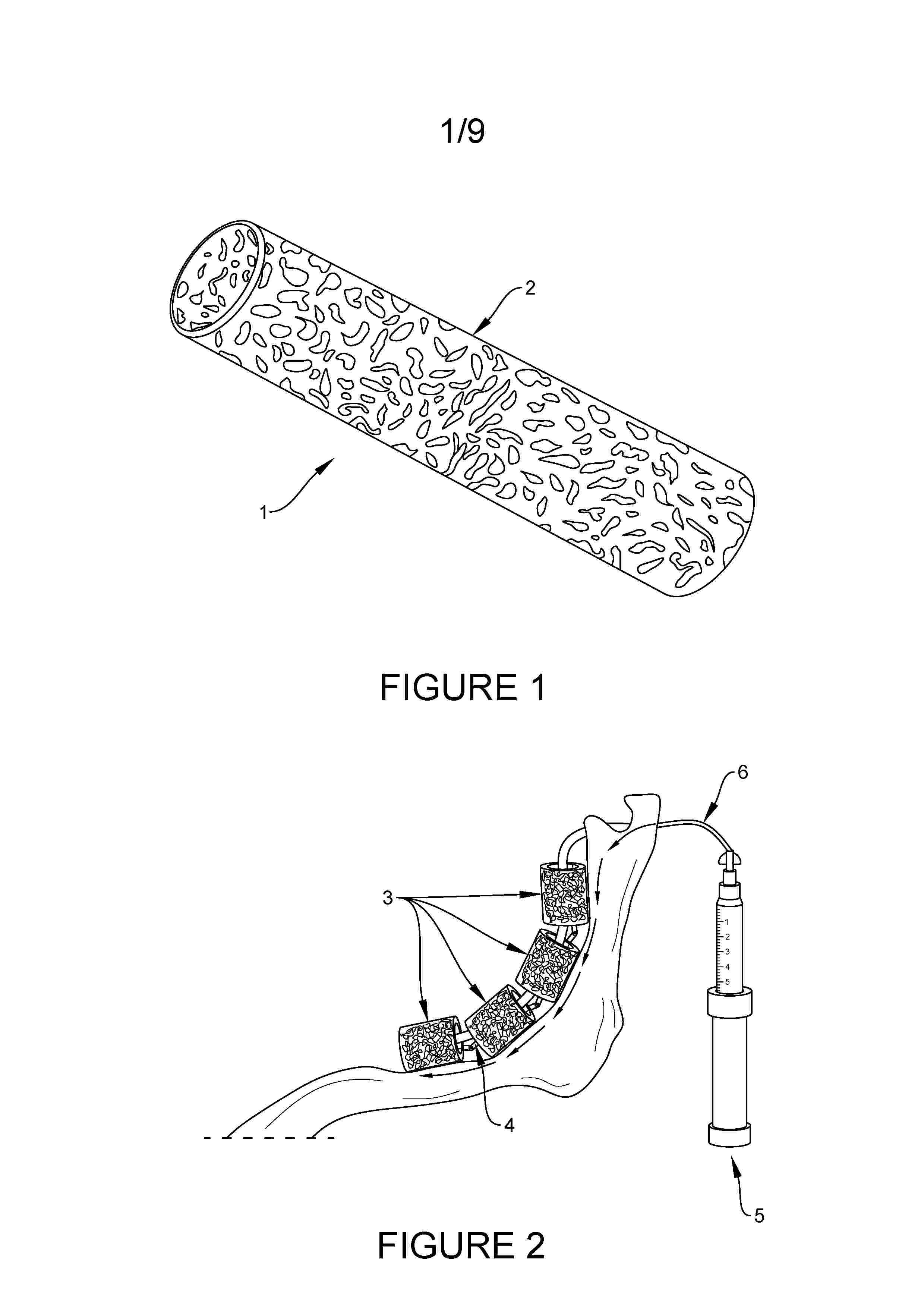
Fixation devices for implants and related methods are generally described. The fixation device may be wrapped around the implant prior to delivery to reduce the risk of implant migration after implantation. The device may have porosity to induce tissue ingrowth around the implant and improve surgical outcome. The device can be used for rapidly preparing implants and can adapt to implants of various sizes. In some embodiments, the device may include a central portion for covering an anterior surface of a breast implant and a peripheral portion for covering a posterior surface of the implant. The peripheral portion, which may include a continuous rim or a plurality of legs, may include a tab that secures the fixation device to an implantation site. In some embodiments, a tether may be coupled to a distal end portion of the peripheral portion, thereby serving as a pull cord that quickly wraps the device around the implant.
Resumen de: AU2024276549A1
Methods of manufacturing a porous device for diffusing a fragrance using additive manufacturing including iteratively spreading a loose powder and selecting at least one process parameter to create a porosity within the porous device adapted to carry the fragrance from a fragrance reservoir to an exterior surface of the porous device. An additively manufactured device for diffusing a fragrance includes a diffuser having a plurality of pores defining a porosity of the diffuser, the porosity defining at least one of a rate of absorption, an amount of storage, and a rate of diffusion of the fragrance. A method of using a porous device includes filling the plurality of pores with fragrance and diffusing the fragrance from an exterior surface.
Resumen de: CN121127203A
A computer-implemented method for predicting tooth positions, comprising: a) defining a plurality of tooth positions corresponding to a plurality of teeth in a dental arch; b) defining a plurality of appliance positions of the orthodontic appliance; c) determining one or more tooth position differences for each tooth; d) determining one or more appliance position differences for each tooth; e) determining one or more appliance-tooth relative differences for each tooth; f) modifying each tooth position of the plurality of tooth positions based on the corresponding one or more appliance-tooth relative differences; g) iteratively repeating steps c) to f) until a maximum change in each of the plurality of tooth positions is less than a convergence threshold; and h) defining the plurality of tooth positions obtained in the final iteration of step g) as a plurality of predicted tooth positions.
Resumen de: GB2644212A
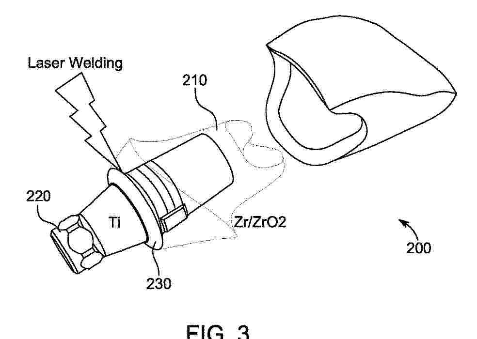
A method of manufacturing a dental implant device 200 comprising affixing a first 210 and second 220 implant member using laser welding, joining or soldering such that there’s no gap. The first implant member is at least partially formed of a ceramic material and the second implant member is at least partially formed of a metal. The implant member comprises melting at least part of the material of the first and second implant member at the same time using a two-wavelength laser. Wherein the first wavelength is selected to be adsorbed by the first material and the second wavelength is selected to be adsorbed by the second implant. An associated dental implant is also provided. Additional methods are also provided which may comprise the use of infra or near infra red lasers, depositing metal on the implant members, positioning biocompatible soldering material in the vicinity of the first and second implant member and forming, using a 3d printer, the first and second implant member using a first and second powder material respectively.
Resumen de: GB2700936A
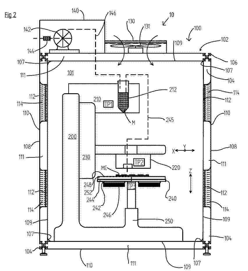
A controller (300, figure 4) for a 3D bioprinter 10, the controller being operable to control the functioning of the 3D bioprinter housed within an internal area of an enclosure 100, to produce a 3D printed construct in accordance with a 3D printing file, said controller being further operable to control the temperature of at least two temperature regulating devices 112, 242, by means of control inputs from plural temperature sensors TP1,TP2,TP3, at least one sensor sensing the temperature of the print bed 240, of a print material store 210, and/or at least one temperature sensor of the bioprinter head 220, and wherein said temperature regulation includes causing: heating of one or more of said at least two devices, cooling of one or more of said devices, or heating of one of the devices at the same time as cooling of another of the devices. The temperature control may be a proportional-integral-derivative (PID) routine or circuit. A 3D printing assembly is also provided, suitable for bioprinting, including an enclosure defining an internal area 101 and one or more bioink material vial(s) 212 selectively fluidically interconnectable to the print head. Figure 2
Resumen de: US20260069422A1
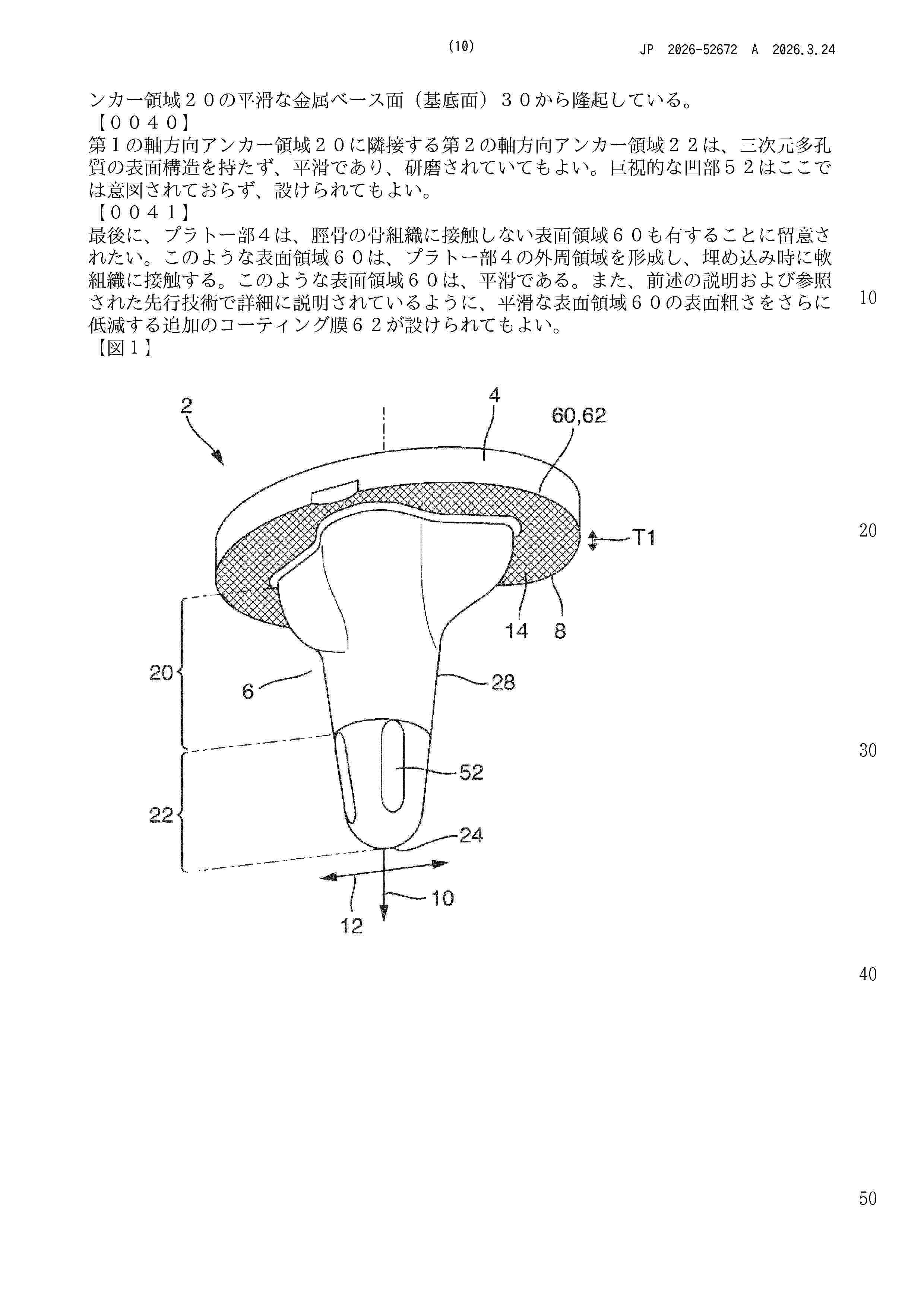
A tibia implant for joint replacement has a plateau section and an anchor section projecting from a tibia-facing side of the plateau section. The anchor section is insertable into a channel in a tibia bone. The plateau section has a first surface structure that is a porous open-pore surface structure with bridges, webs or wall regions that can be gripped from behind in the axial direction on the tibia-facing side that comes into contact with tibia bone tissue. The open-pore surface structure has a first roughness. The anchor section is connected to the plateau section in a first axial anchor region on the circumferential side has a surface structure with a second roughness that is lower than the first roughness. The anchor section has a second axially free-ending axial anchor region with a smooth surface in the axial direction adjacent to the first axial anchor region.
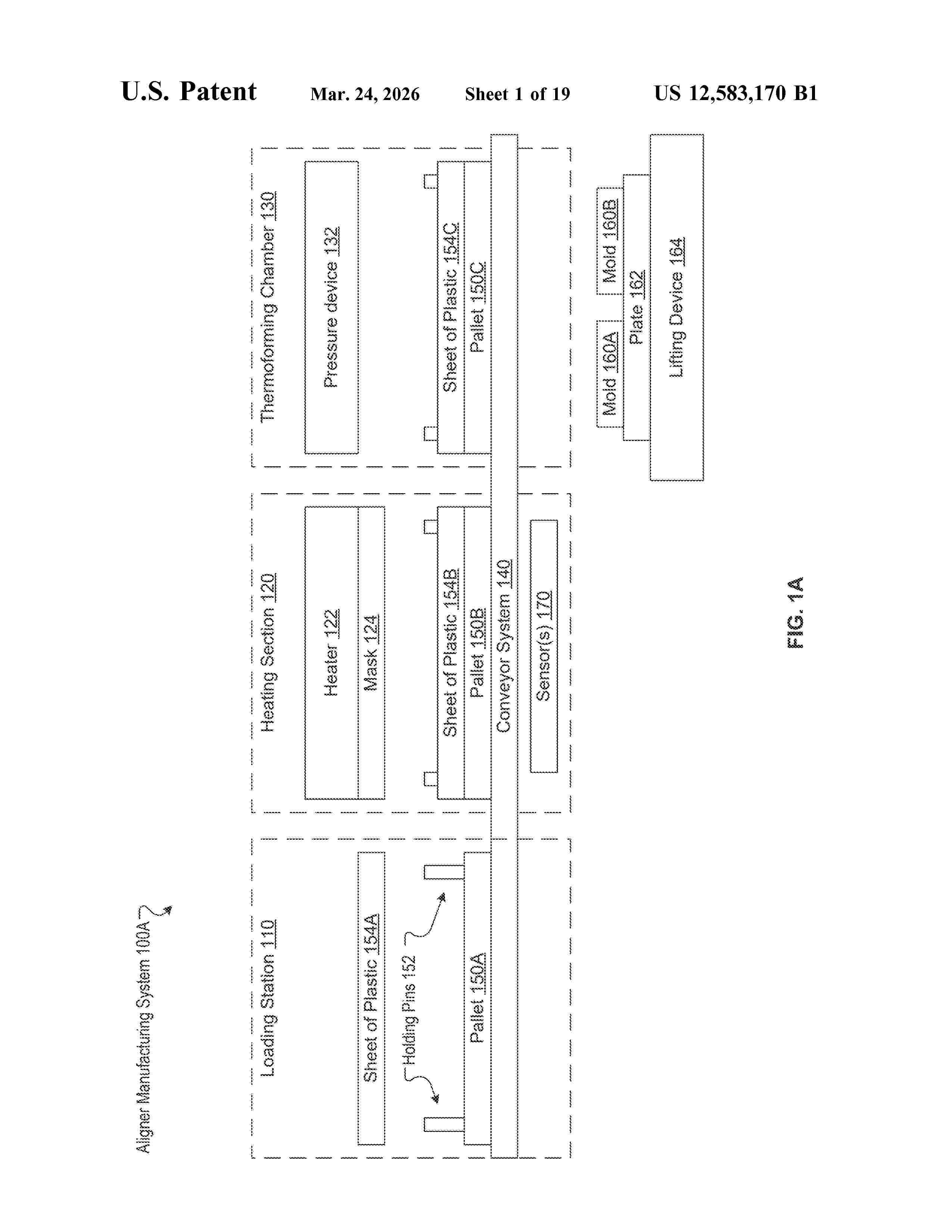
Resumen de: US12583170B1
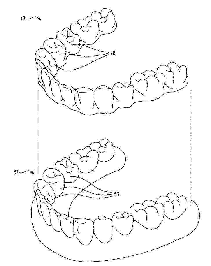
A method includes: receiving first and second digital models associated with a dental arch of a patient corresponding to first and second stages of orthodontic treatment; printing a first mold of a first dental arch based on the first digital model and a second mold of a second dental arch based on the second digital model; securing the first and second molds to a plate; heating a sheet of plastic to generate a heated sheet, and surrounding the sheet of plastic with a mask during the heating of the sheet of plastic to minimize heat transfer to other sheets of plastic; and simultaneously thermoforming the heated sheet over the first and second molds to form a first aligner shaped to fit the first dental arch and a second aligner shaped to fit the second dental arch.
Resumen de: US20260013994A1
Implantable devices for spinal cord and peripheral nerve injury are described. The implants include a three-dimensional printed structure having stem cells disposed therein. Also disclosed are methods of treating neuronal injuries with the disclosed implants.
Resumen de: CN121714758A
本发明公开了一种时空响应性多功能3D打印骨修复支架及其制备方法和应用。将PDGF‑BB负载于快释载体,MgP负载于慢释载体,通过3D打印制备出内层负载MgP、外层负载PDGF‑BB的多功能3D打印骨修复支架。其中PDGF‑BB作为免疫调节剂,MgP作为再生诱导剂,将其植入体内后可根据载体降解速率的不同,时空响应性先降解释放PDGF‑BB实现早期免疫调节和内皮细胞募集,后降解释放镁离子和磷酸根离子与PDGF‑BB协同促进中后期血管化和成骨。经体外体内实验证实,本发明提供的3D打印骨修复支架兼具免疫调节、促进血管生成和成骨性能,能够用于炎症性骨损伤修复,尤其是适用于糖尿病引起的炎症性骨损伤修复。
Resumen de: CN121718752A
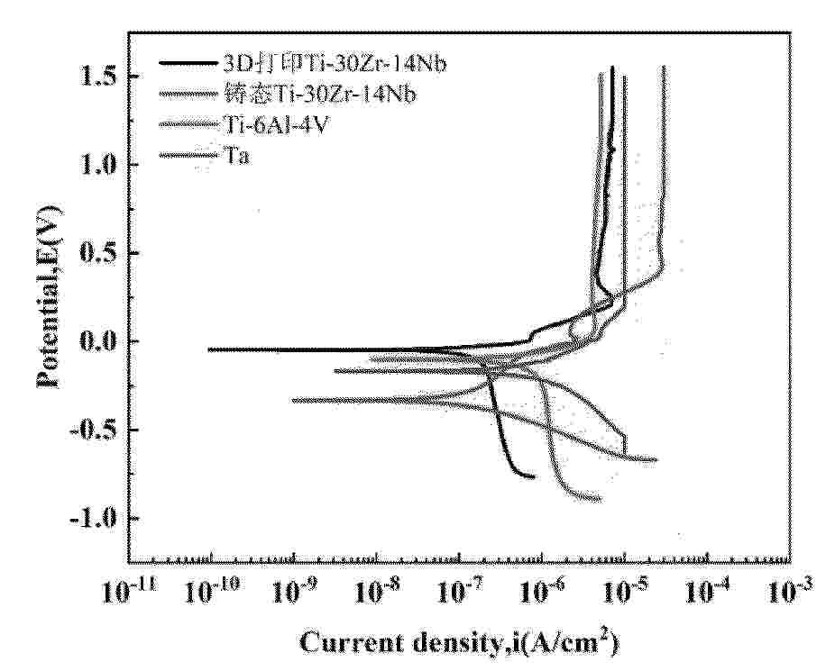
本发明公开了一种用于骨植入的Ti‑Zr‑Nb中熵合金多孔构件及制备和应用。该多孔构件的各元素的原子百分比为:Ti 52‑57 at.%,Zr 27‑32 at.%,Nb 12‑16 at.%,以不可避免的杂质元素;且多孔构件的结构由相互连通的大孔和小孔构成,孔隙率为 35%‑45%。屈服强度为 127‑216 MPa,弹性模量为 5.0‑8.5 GPa,且所述大孔孔径为 550‑700μm;所述小孔孔径为 200‑400μm,且大孔与小孔的连通率≥90%。该多孔构件在模拟体液中表现出优异的耐腐蚀性(自腐蚀电流密度 0.22×10⁻6 A/cm2),且能支持骨髓间充质干细胞的黏附、增殖与成骨分化,生物相容性良好。本发明为高性能、个性化骨植入物的制备提供了新的材料体系和成形工艺,具有重要的临床应用价值。
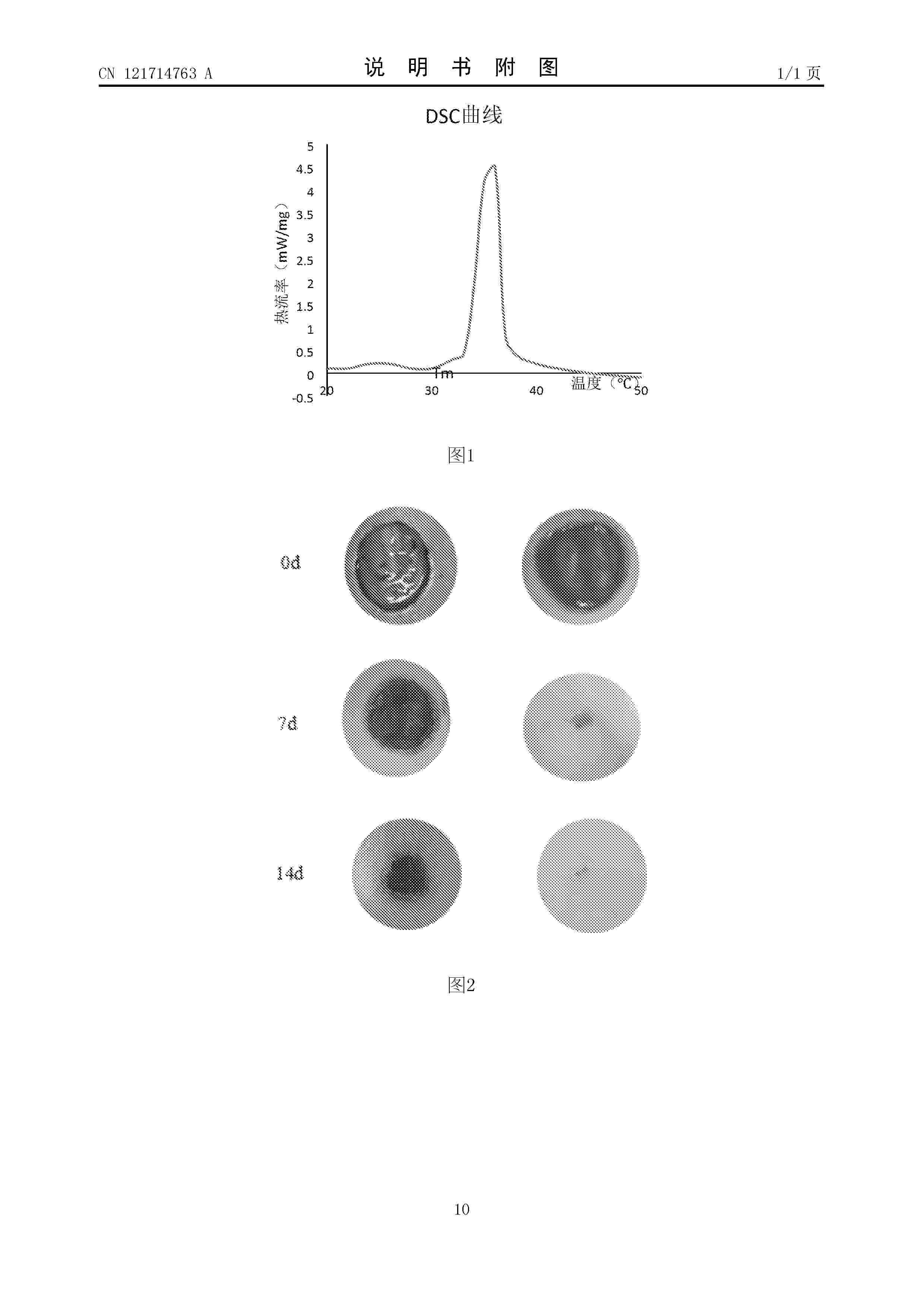
Resumen de: CN121714763A
本发明涉及一种生物墨水、4D生物活性支架及其制备方法与应用,属于生物技术领域。本发明提供的生物墨水包括重组贻贝粘蛋白、表皮生长因子(EGF)、甲基丙烯酸酐化明胶、透明质酸、聚多巴胺、N‑异丙基丙烯酰胺、海藻糖、四丁基溴化铵、LAP和18‑冠醚‑6。本发明的生物墨水用于制备4D生物活性支架,得到的支架具有良好的细胞粘附作用,能按需释放活性因子,同时具有温度响应性,能在37℃体温触发变形,实现对组织的快速修复,且安全性良好,可应用于组织再生与医疗美容修复。
Resumen de: US2025367422A1
A microneedle array is provided for administrating a drug or other substance into a biological tissue. The array includes a base substrate; a primary funnel portion extending from one side of the base substrate; and two or more solid microneedles extending from the primary funnel portion, wherein the two or more microneedles comprise the substance of interest. Methods for making an array of microneedles are also provided. The method may include providing a non-porous and gas-permeable mold having a two or more cavities each of which defines a microneedle; filling the cavities with a fluid material which includes a substance of interest and a liquid vehicle; drying the fluid material to remove at least a portion of the liquid vehicle and form a plurality of microneedles that include the substance of interest, wherein the filling is conducted with a pressure differential applied between opposed surfaces of the mold.
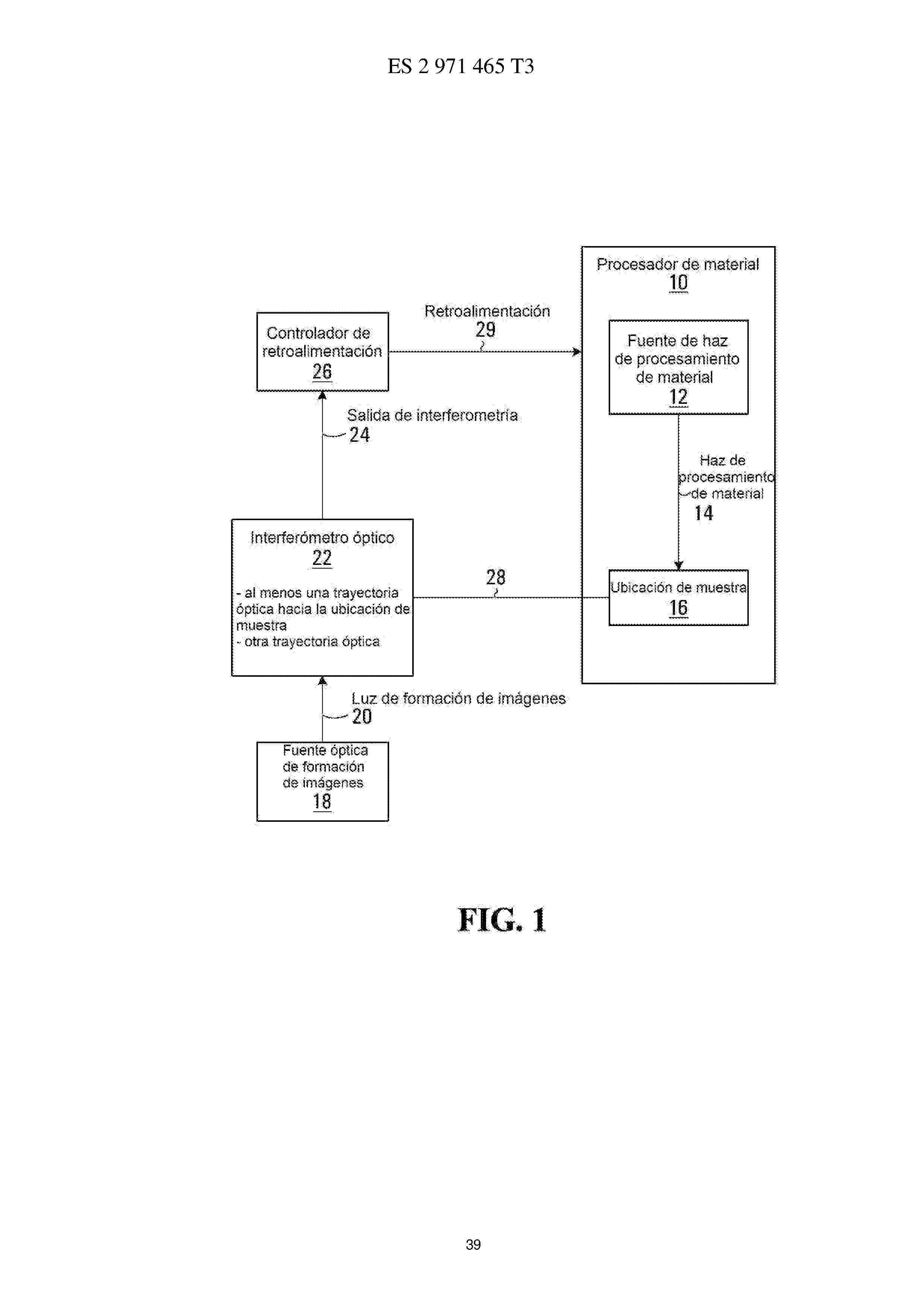
Resumen de: EP4306235A2
Methods and systems are provided for using optical interferometry in the context of material modification processes such as surgical laser or welding applications. An imaging optical source that produces imaging light. A feedback controller controls at least one processing parameter of the material modification process based on an interferometry output generated using the imaging light. A method of processing interferograms is provided based on homodyne filtering. A method of generating a record of a material modification process using an interferometry output is provided.
Resumen de: CN121175081A
A method of modifying a surface of a three-dimensional (3D) article includes immersing at least a portion of the 3D article in a buffer solution of a functionalized peptide such that the functionalized peptide reacts with a reactive group on the surface of the 3D article; and washing at least a portion of the 3D article to remove unreacted functionalized peptides. The surface-modified 3D article comprises a plurality of peptides covalently bonded to a surface of the 3D article by cysteine bridging. The surface-modified 3D articles can be used as scaffolds for constructing biological tissues or implants.
Resumen de: CN121697158A
本发明公开了应用于肱骨近端骨折的解剖型骨水泥棒支撑的倒模,属于肱骨近端骨折支撑技术领域,该应用于肱骨近端骨折的解剖型骨水泥棒支撑的倒模,包括用于合模成型的左模具和右模具,倒模还包括:左模具内部成型面开设有相贯通的左型芯上模腔和左型芯下模腔,右模具内部成型面开设有右型芯模腔;左模具和右模具精准对接合模制备适配肱骨近端髓内解剖结构的支撑棒;通过左模具和右模具能够快速制备骨水泥支撑棒,操作快速便捷,并且骨水泥支撑棒成型的形态和大小稳定,符合手术需要,不需要反复调整修正,并且左模具和右模具能够通过3D打印技术制备或者采用金属材料制备,制备的模具能够重复利用,辅助手术操作。
Resumen de: CN120678548A
The invention discloses a complete denture and a complete-process digital complete denture manufacturing method. A traditional impression is replaced by an optical impression, mechanical face arch and Gothic arch tracing is replaced by electronic motion face arch and mandibular motion track tracing, traditional jaw position recording is replaced by combining mandibular track characteristics displayed by a voice method, the technical sensitivity is lower, and jaw position relation recording is more accurate; after mouth scanning, an occlusion scheme is directly generated through software tooth pre-arrangement and an electronic face arch, the middle steps of wax pattern try-on, traditional jaw position recording and the like are omitted, digital manufacturing is adopted, manual operation is reduced, false teeth are directly output through the three-dimensional printing/cutting technology, the tedious procedures of boxing, tooth boiling, grinding and the like are omitted, the manufacturing period is greatly shortened, and the manufacturing cost is reduced. And the subsequent secondary copying or remanufacturing time and the adaptation time are greatly shortened, a digital solution is provided for reconstructing the jaw position through full-mouth planting occlusion, and development, integration and the like of digital and intelligent data are facilitated.
Resumen de: CN121697214A
本发明提供了一种利用DLP光固化3D打印实现颜色连续梯度过渡的方法,属于增材制造技术领域。步骤包括:提供双波长DLP光固化打印设备,调整打印焦平面,对三维数字模型进行分割与切片处理,设定双波长曝光参数,制备含pH响应染料、光酸发生器等组分的光敏树脂浆料,通过可见光固化成型与紫外光调控颜色的往复动态切换完成逐层打印,经后处理得到目标制件。本发明方法通过单光机双波长(365nm紫外光与460nm可见光)超高速动态切换,结合特定配方的光敏树脂浆料,实现多色材料的高分辨率、连续渐变一体化成形。
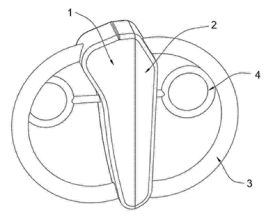
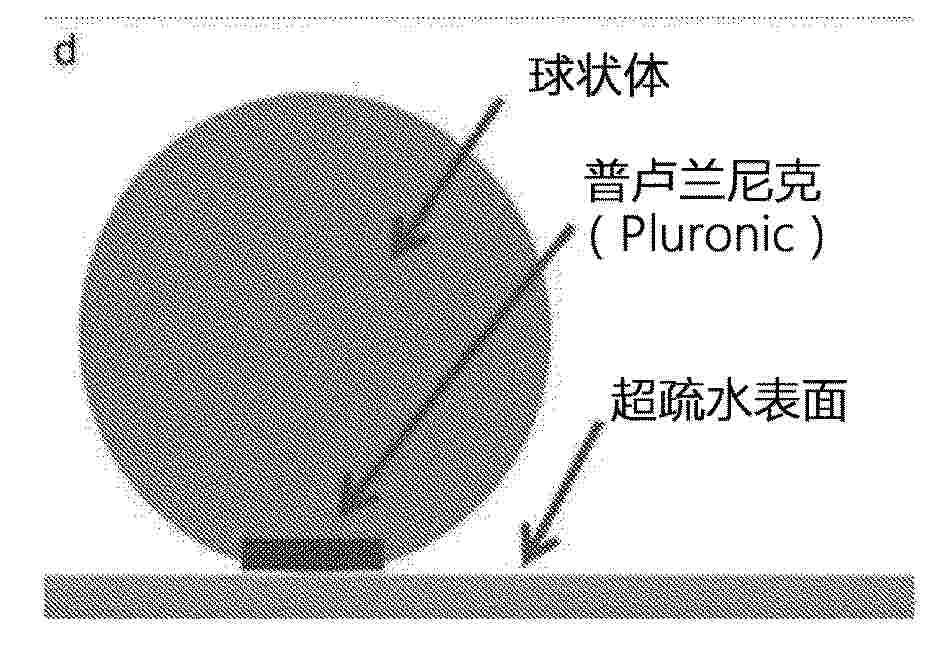
Nº publicación: CN121695334A 20/03/2026
Solicitante:
路易斯维尔大学研究基金会
Resumen de: US2019381213A1
A method of making a spheroid is provided that includes the step of providing a suspension having one or more biologically-relevant materials dispersed within a biocompatible medium. An amount of a hydrophilic material is deposited on a defined area of a super-hydrophobic surface, and a droplet of the suspension is bioprinted onto the hydrophilic material positioned on the super-hydrophobic to thereby create the spheroid.
 BOPI
BOPI
 Sede Electrónica