Si deseas distinguir tus productos, servicios o ambos de los de otra empresa, es posible que necesites una marca o nombre comercial. Descubre qué son, en qué consiste su procedimiento de registro y qué implica.

Información sobre los plazos de presentación de solicitudes de transformación de marcas de la Unión Europea en marca nacional española. Más información


Si tienes un nuevo dispositivo, producto o procedimiento que resuelva un problema técnico o tenga una ventaja práctica, existen distintas formas de protegerlo en España y en otros países. Descubre cómo hacerlo.

¿Tu innovación reside en la estética, la ornamentación o la apariencia de tu producto? Protégela mediante un diseño industrial. Descubre qué derechos confiere el registro y cómo realizar la tramitación.

Las indicaciones geográficas protegen el nombre de un producto originario de una zona geográfica, a la cual le debe una determinada calidad, reputación u otra característica. Descubre qué son, en qué consiste su procedimiento de registro y qué beneficios conceden.

Las patentes publicadas en todo el mundo son una valiosa fuente de información científica, técnica y comercial.

Si eres emprendedor/a o una empresa y quieres potenciar y mejorar la rentabilidad de tu negocio protegiendo de forma adecuada los activos intangibles de tu organización, en este espacio encontrarás lo necesario.


 374
resultados
374
resultados
 Última actualización
26/08/2026 [07:09:00]
Última actualización
26/08/2026 [07:09:00]


 Resultados 100 a 125 de 374

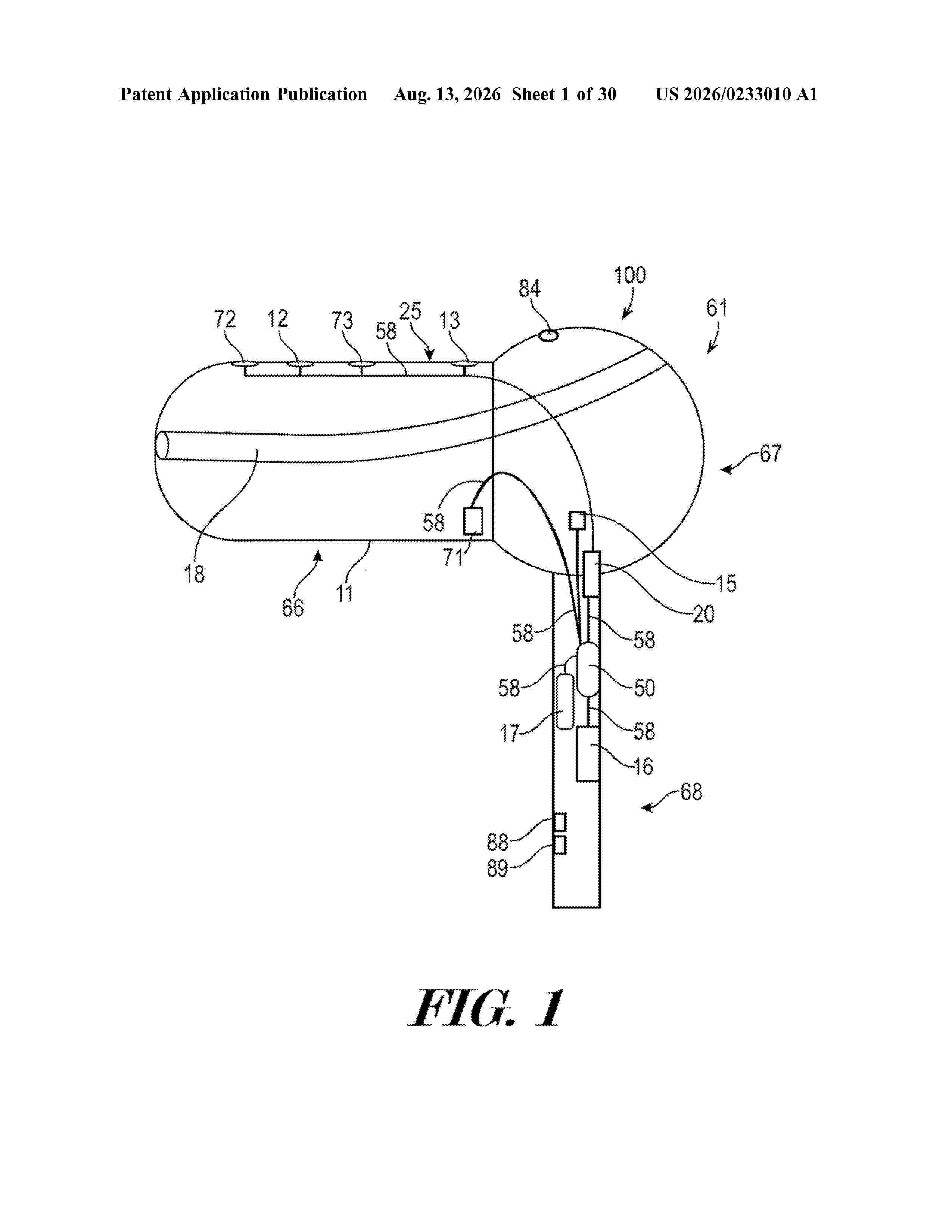
Resumen de: US20260233010A1
0000 An extended auricular electroencephalogram (EEG) monitoring system has a processing unit configured to analyze EEG data to detect neuropsychiatric disorders and impending neuropsychiatric disorders. An automatic detection-therapy system includes the auricular EEG monitoring system and one or more neuromodulation units. When presence of EEG signals suggestive of a neuropsychiatric disorder or an impending neuropsychiatric disorder is detected by the processing unit, the neuromodulation unit is configured to immediately start neuromodulating electric stimulation automatically to one or various combinations of the following: auricular branch of vagus nerve, supraorbital nerve, infraorbital nerve, auriculotemporal nerve, occipital nerve and greater auricular nerve to alleviate or abort the condition. A combined neuromodulation system having two or three neuromodulating components with synergistic effect is useful for health maintenance and prophylaxis of neuropsychiatric disorders. These earbud-shaped systems, with automated setup designs, are wearable, user-installable, user-removable and freely ambulatory.

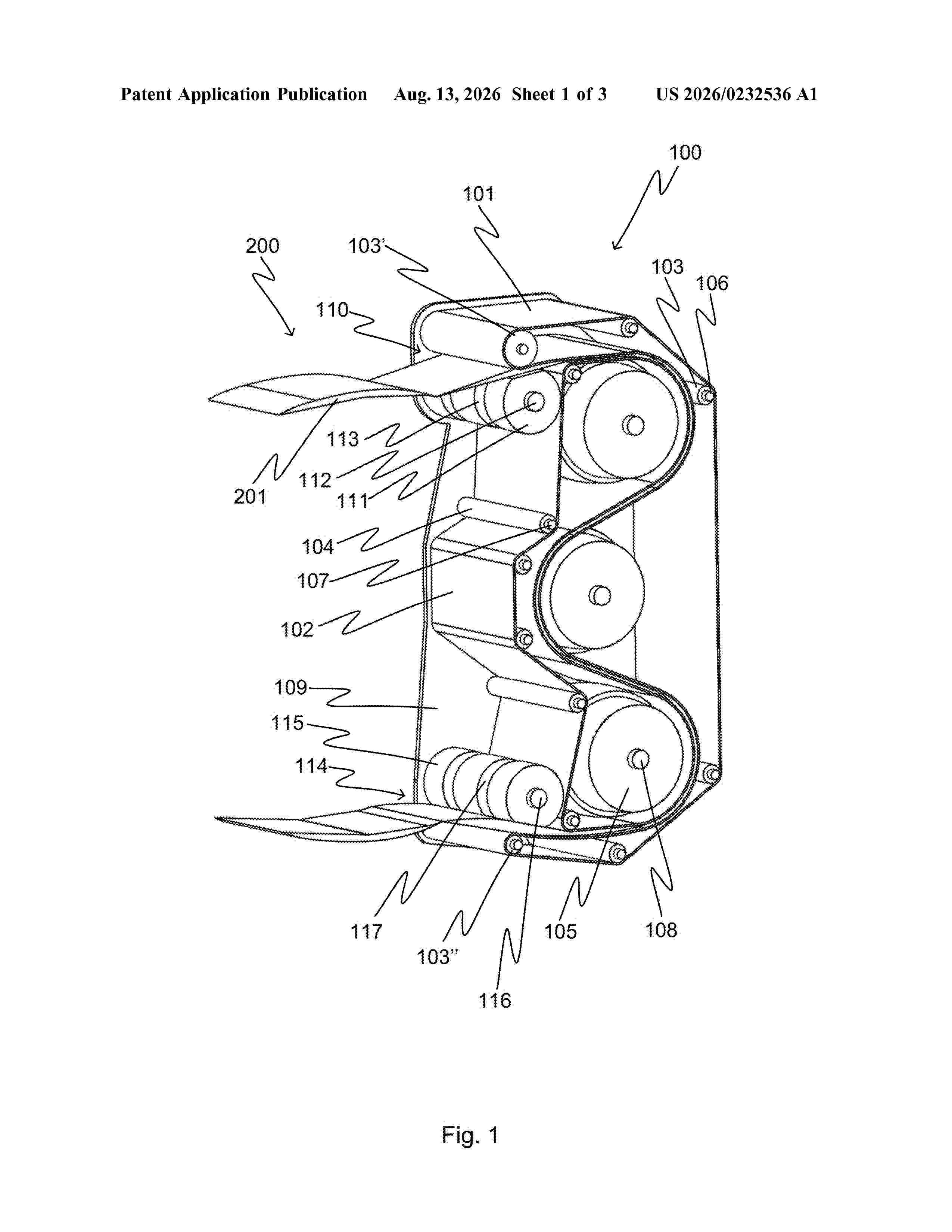
Resumen de: US20260232536A1
A device conveys medication sachets. The device includes an inlet for receiving the medication sachets, an outlet for discharging the conveyed medication sachets, a first endless belt, first guide elements arranged to guide the first endless belt along a first path. First guide elements include a first guide roller, a second endless belt, a plurality of second guide elements arranged to guide the second endless belt along a second path. The second guide elements include a second guide roller, a plurality of third guide rollers arranged to guide the first endless belt and the second endless belt along a third path. The medication sachets are conveyed between the first endless belt and the second endless belt along the third path from the inlet to the outlet. A device rotates at least one of the first, second or third guide rollers. A medication dispenser dispenses the medication sachets.
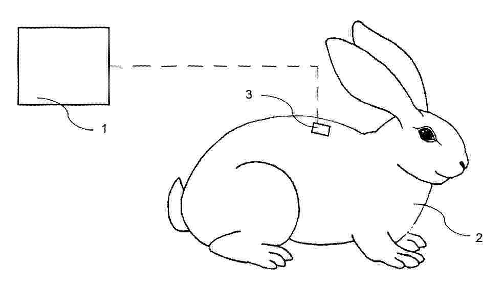
Resumen de: DE102025104748A1
Die vorliegende Erfindung betrifft ein Sensorsystem zur Überwachung von Tieren, insbesondere von Kleintieren, umfassend: eine Stromversorgungs- und/oder eine Stromerzeugungsquelle, die das Sensorsystem mit Strom versorgt; einen loT-Sensor, der Erfassungsdaten für das Sensorsystem bereitstellt, wobei der loT-Sensor dazu eingerichtet und dafür vorgesehen ist, auf der Haut, dem Fell oder unter die Haut eines Tieres transplantiert zu werden; und einen Beschleunigungsmesser oder einen GPS fähigen Sensor, der Bewegungen des Sensorsystems erkennt, um Bewegungsdaten des loT-Sensors zu erhalten; einen Transceiver, der mit dem loT-Sensor in einem loT-System kommuniziert; einen Prozessor, der: die Bewegungsdaten vom loT-Sensor empfängt und eine Aktivierung des loT-Sensors und eine Kommunikationsfrequenz des Transceivers mit dem loT-Sensor steuert; und eine loT-Verbindung, die einen Zugangspunkt mit dem loT-Sensor verbindet, wobei der Transceiver eine Antenneneinrichtung umfasst, und diese Antenne auf einer Sensorplatine verläuft und/oder die Antenne von dem Transceiver und dem loT-Sensor weg entlang und innerhalb einer Körpermulde und/oder Körperöffnung des Tieres geführt ist und dabei die Antenne mit einem Material gebildet ist, welches einen Biegeradius von zumindest einem Millimeter zerstörungsfrei und reversibel standhält und sich damit an den Oberflächenverlauf des Tieres oder, wenn unter die Haut implantiert, an den unter der Haut verlaufenden Knochen und/oder Weichte
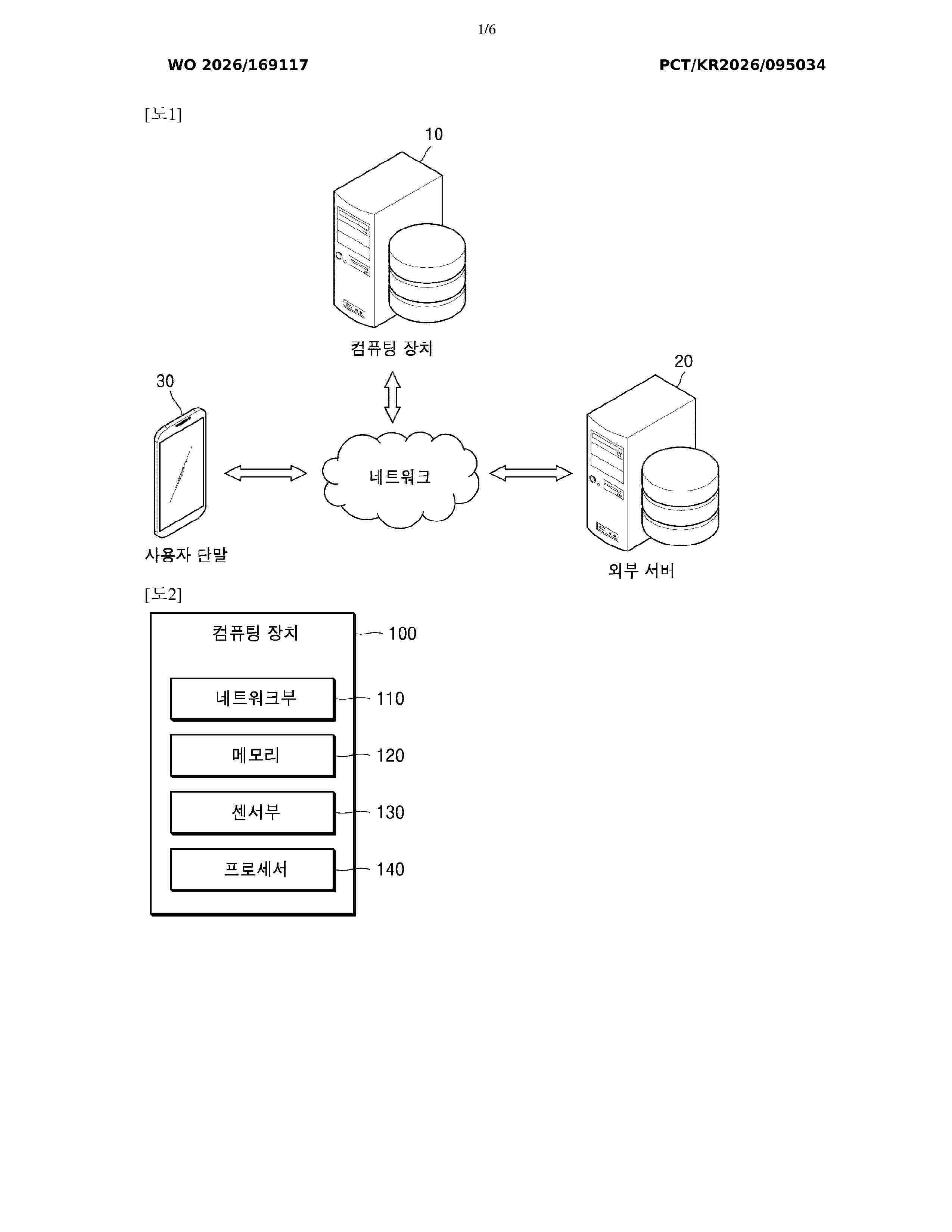
Resumen de: WO2026169117A1
The present disclosure relates to a method and a device for generating a digital electrocardiogram signal on the basis of an electrocardiogram record. According to an embodiment of the present disclosure, a method for generating a digital electrocardiogram signal on the basis of an electrocardiogram record is provided, the method including the steps of: performing preprocess to extract first electrocardiogram information on the basis of a first electrocardiogram record; training an artificial intelligence segmentation model on the basis of the first electrocardiogram record and the first electrocardiogram information; segmenting a second electrocardiogram record into second electrocardiogram information by using the second electrocardiogram record as an input of the trained artificial intelligence segmentation model; and converting the segmented second electrocardiogram information into a digital electrocardiogram signal.
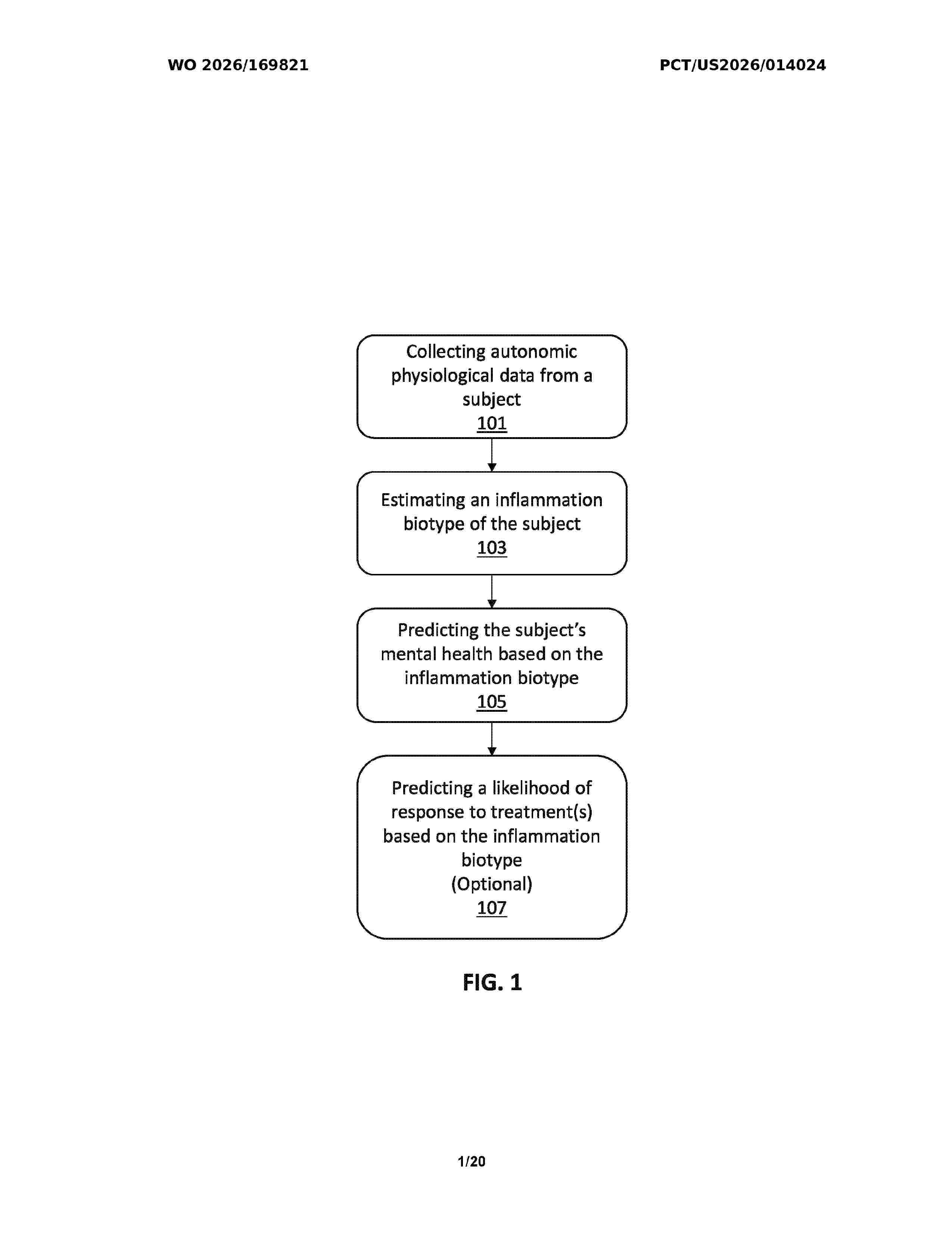
Resumen de: WO2026169821A1
Systems and methods for assessing inflammatory response and mental health of subjects. The assessments may be based on measurements taken non-invasively. Methods may include classifying an inflammation risk for the subject based on physiological data and estimating a mental health status of the subject. The inflammation risk classification may be used to predict a likelihood of the subject's response to anti-inflammatory treatment. The systems and methods may include the use of machine learning models.
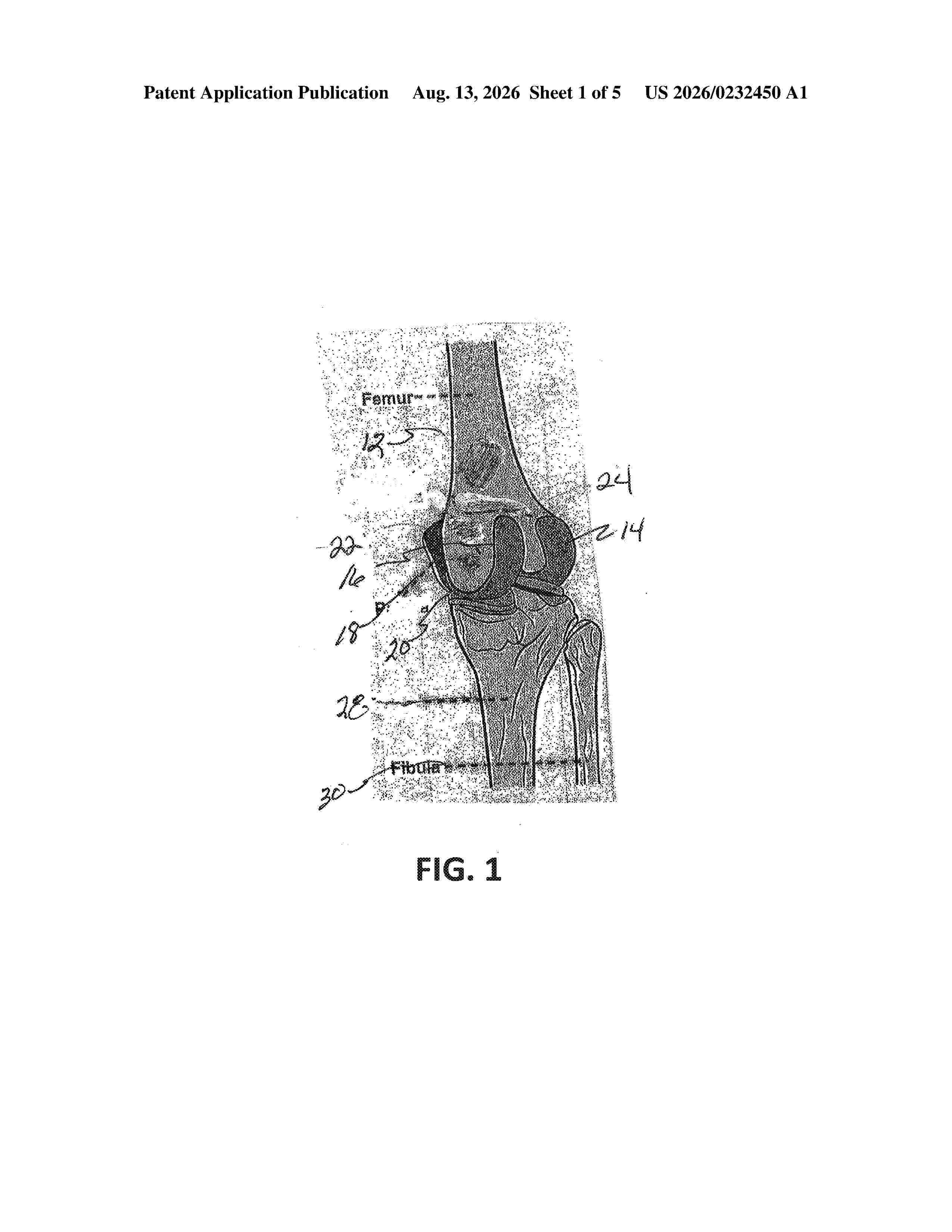
Resumen de: US20260232450A1
0000 An additive manufactured knee femoral component replacement for either a full or partial replacement has an outer wall formed from a flexible polymer and a structural body or medial portion formed from a fiber reinforced polymer. The replacement can have a single posterior condyle or a pair of spaced apart condyles with a gap therebetween to accommodate ligaments extending therethrough.
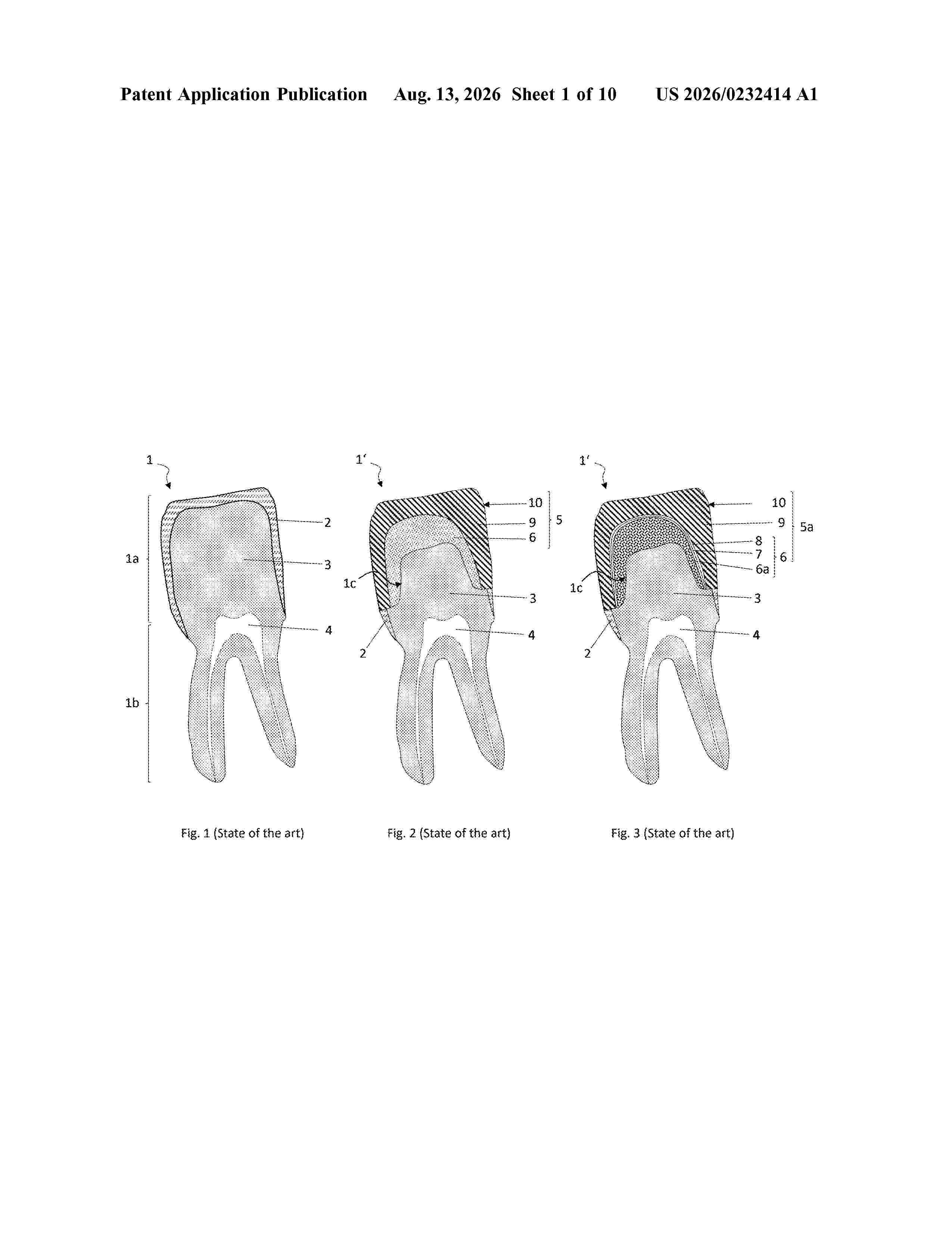
Resumen de: US20260232414A1
0000 A method and a system for the automated, software-based manufacturing of a veneer of a fixed dental prosthesis by coating a dental preform with at least one compound forming a ceramic paste that reproduces the dentin of a natural tooth. The preform is coated based on a 3D layer structure in which each layer is divided into volume elements. Each volume element is filled with a portion of the ceramic paste by aligning the preform with a manipulator so that a ground surface of the volume element to be filled, at an application point on the ground surface, is oriented substantially perpendicular to an ejection direction with the application point lying on the ejection direction. A print head applies the respective portion of the paste by jerking a valve element so that the portion is formed and projected in a coherent manner toward the application point and fills the volume element in a punctiform manner. The compound used is a ceramic paste formed from a powder of a feldspar ceramic finely dispersed in a solvent and including amounts of ammonium polyacrylate, ethoxylated castor oil and traces of urea.
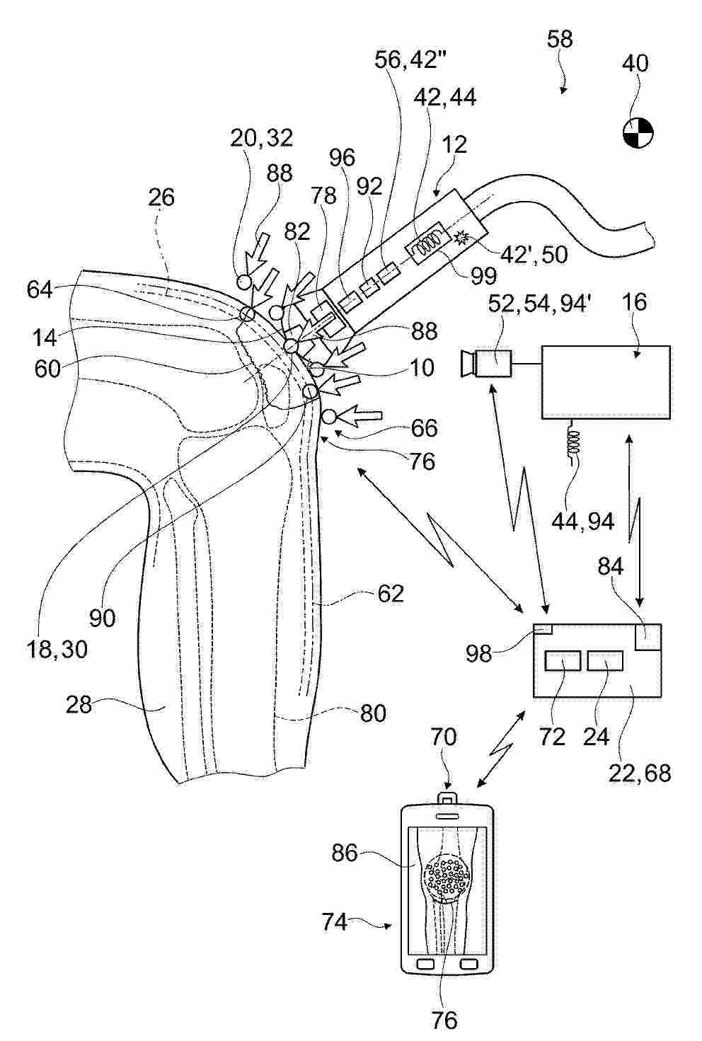
Resumen de: DE102025104781A1
Die Erfindung geht aus von einem Therapiesystem (58) zu einer nicht-chirurgischen Behandlung zumindest eines flächigen Körperbereichs (10) eines Lebewesens, mit einer Applikatoreinheit (12), aufweisend einen Therapiekopf (14) zu einer Applizierung propagierender physikalischer therapeutisch wirksamer Wellen an dem flächigen Körperbereich (10), und ein Trackingsystem (16), welches dazu vorgesehen ist, während mehrerer Behandlungsabschnitte (18, 20) der Behandlung des flächigen Körperbereichs (10) momentane 3D-Positionsdaten (30, 32) der Applikatoreinheit (12) zu ermitteln.Es wird vorgeschlagen, dass das Therapiesystem (58) eine Recheneinheit (22) mit Zugriff auf ein 3D-Ausgangsmodell (26) eines Körperteils (28) eines Lebewesens, aufweist, wobei die Recheneinheit (22) dazu vorgesehen ist, die von der Trackingsystem (16) erfassten 3D-Positionsdaten (30, 32) der Applikatoreinheit (12) zu empfangen, und basierend auf den empfangenen 3D-Positionsdaten (30, 32) eine von dem 3D-Ausgangsmodell (26) ausgehende Oberflächenapproximation (34) des Körperteils (28) mit dem der Behandlung unterzogenen flächigen Körperbereich (10) zu berechnen, und die zu einzelnen Behandlungsabschnitten (18, 20) der Behandlung zugehörigen 3D-Positionsdaten (30, 32) Oberflächenpunkten (36, 38) der Oberflächenapproximation (34) zuzuordnen
Resumen de: WO2026169805A1
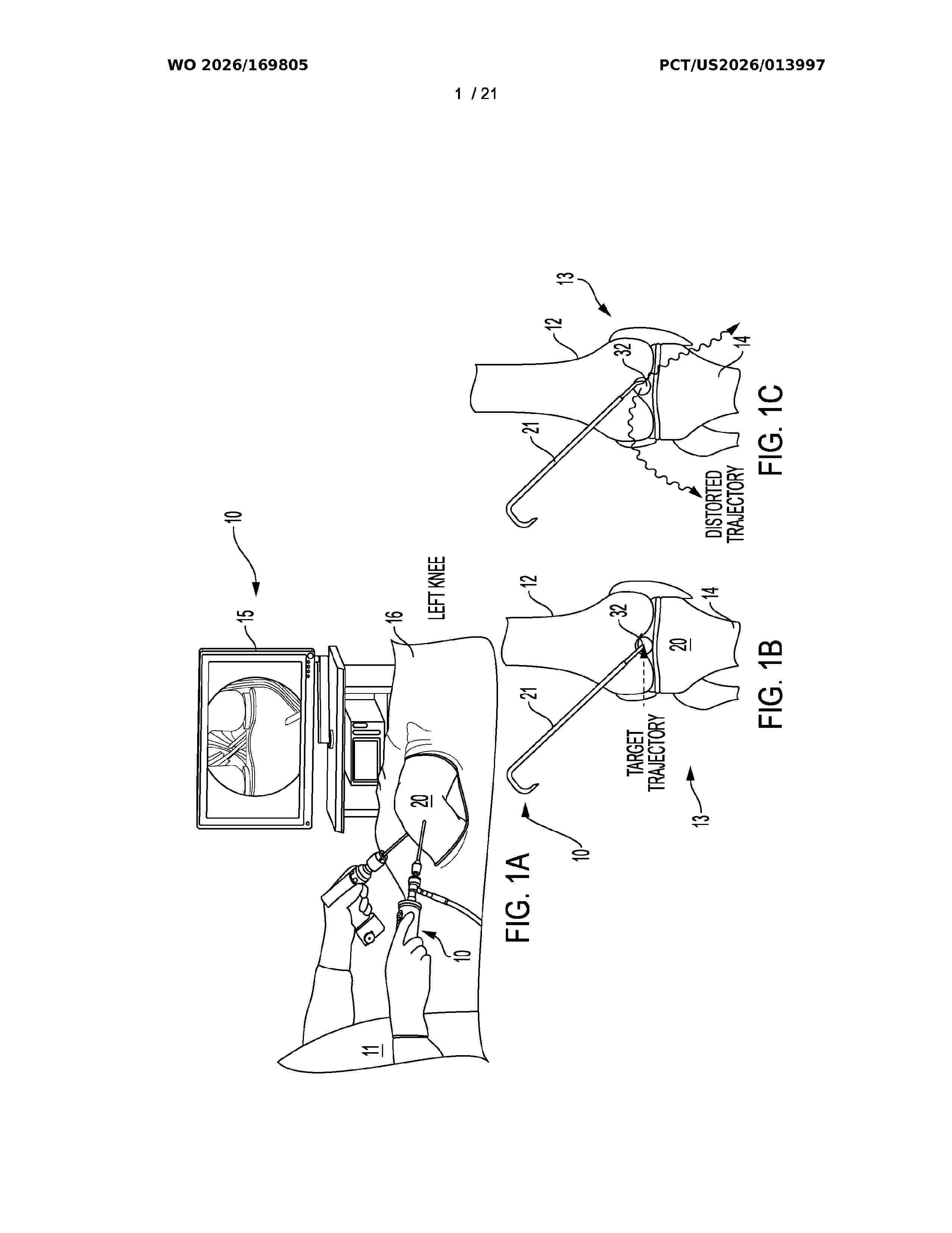
A navigation device, adapter, and system enables real-time three-dimensional depth sensing through standard endoscopic and borescopic instruments, including laparoscopes, arthroscopes, cystoscopes, ureteroscopes, bronchoscopes, sinuscopes, and borescopes. A releasable adapter integrates LiDAR or other optical time-of-flight sensors with off-the-shelf endoscopes and surgical cameras to generate sub-millimeter precision surface reconstructions and register those reconstructions to preoperative or intraoperative scans for accurate image-guided procedures, particularly on rigid anatomy. The system supports surgical guidance, augmented reality visualization, distance -based signal normalization, 3D measurement, luminal traversal tracking, and robotic control. Time-of-flight sensing over a selected visible sub-band enables simultaneous acquisition of white-light, fluorescence, and depth data, with calibration workflows for wide field-of-view optics and adaptive software control of focus, illumination, and rendering.
Resumen de: WO2026170003A1
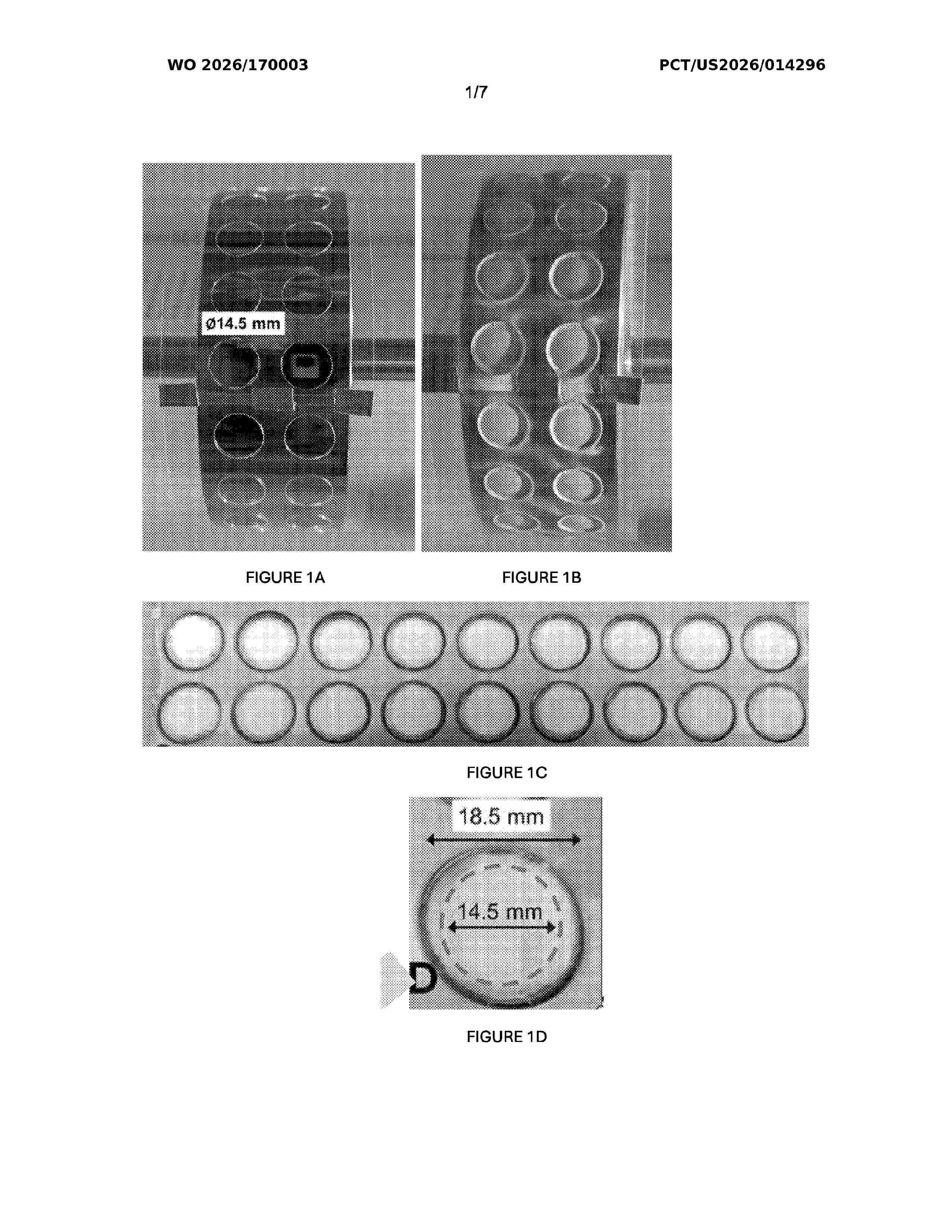
An in vitro 3D hepatocyte model that contains a nanofibrous scaffold designed to mimic the extracellular matrix (ECM) of the human liver, both structurally and biochemically, is described. HepaRG hepatocytes cultured on the 3D scaffold exhibited enhanced metabolic activity compared to traditional 2D cultures, indicating improved hepatocyte functionality. This model offers a physiologically relevant platform for hepatocyte studies, underscoring the model's potential to advance preclinical drug development by replicating liver-specific functions in vitro.
Resumen de: DE102025105136A1
Die Erfindung betrifft ein Verfahren zur Herstellung eines Individual-Zahnimplantats gekennzeichnet durch folgende Schritte:- die Kiefer- und Zahnreihen oder eine Abformung der Kiefer- und Zahnreihen wird digital als erste Information an einem Behandlungsort erfasst;- die Knochen-Hartgewebsstruktur wird zwei- oder dreidimensional geröntgt und digital als zweite Information an dem Behandlungsort erfasst;- die erste Information und die zweite Information werden einem zentralen Datenpool zur Abspeicherung und Bearbeitung in einer Bearbeitungsstation an dem Behandlungsort zugeführt;- auf dem zentralen Datenpool ist eine künstliche Intelligenz (KI) vorhanden, wobei die kI in Abgleich der ersten Information und der zweiten Information eine Fehlsituation ermittelt, und diese Fehlsituation in Abhängigkeit von der ersten Information und der zweiten Information ermittelt und durch das Individual-Zahnimplantat substituiert, wobei eine Form, eine Größe, eine Position und eine Oberflächenstruktur sich an der ersten Information orientiert, geeignet, dass das Individual-Zahnimplantat die Funktion eines natürlichen Zahns oder einer natürlichen Zahnwurzel optimal nachbildet.
Resumen de: US20260232228A1
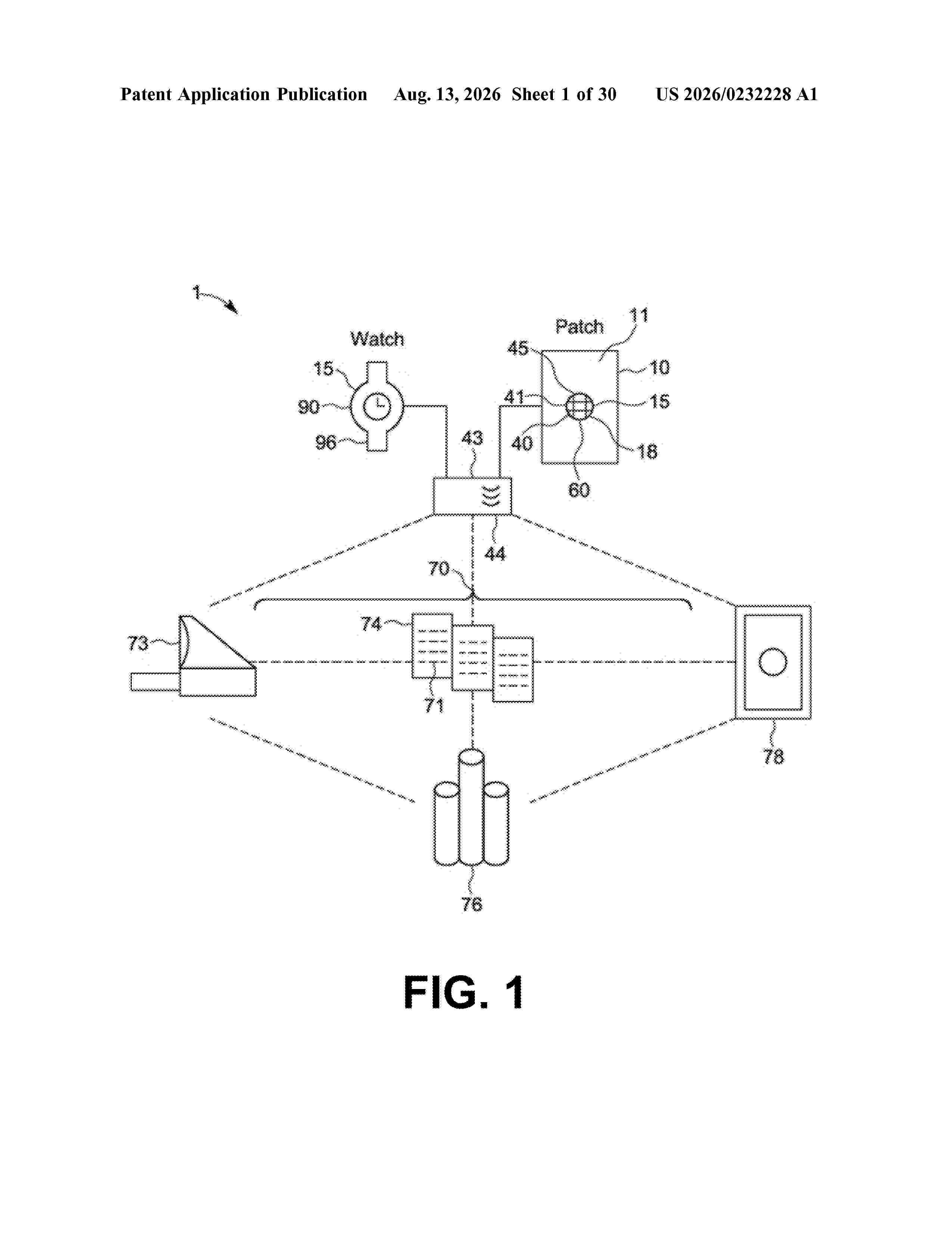
The methods can include receiving calibrating spectral data from a non-invasive measurement device, known measurement data that is indicative of a concentration of a particular biomolecule in the body of the wearer and was obtained contemporaneously with the calibrating spectral data; generating a signature for the wearer by at least determining, via a neural network, one or more correspondences between or among the calibrating spectral data and the known measurement data; and storing the signature. The methods can include receiving present spectral data from the non-invasive measurement device and processing the present spectral data using the signature to estimate a present concentration of the particular biomolecule in the body.
Resumen de: US20260232870A1
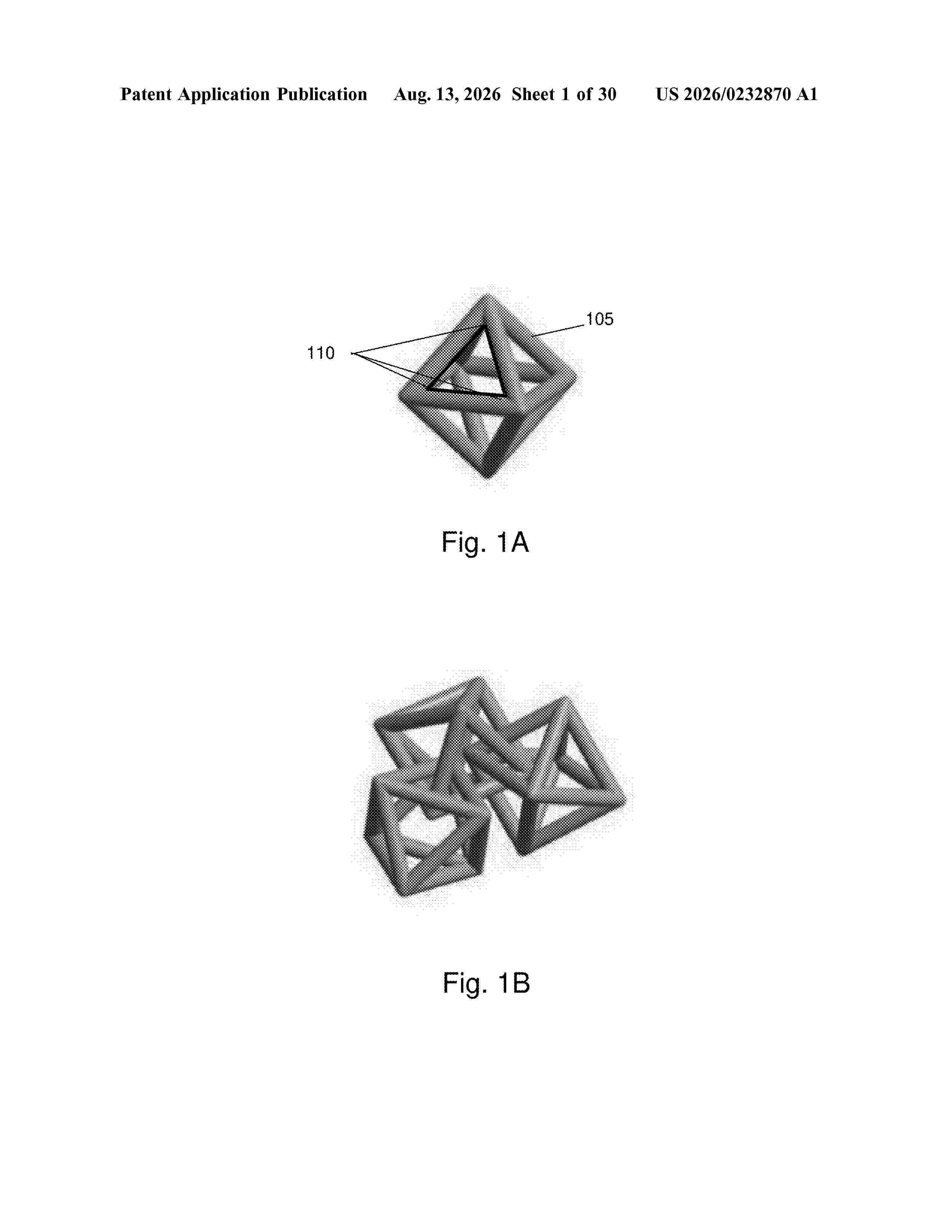
Described are 3D polycatenated architected materials, systems, and methods. The material comprises discrete unit particles mechanically interlocked to form a 3D network patterned after a crystalline topology. This structure provides a tunable dual mechanical response, transitioning from a fluid-like regime to a solid-like regime at a programmable “critical jamming strain.” Methods are disclosed for designing the material using a closed-loop digital twin, manufacturing the material via additive processes, and preconditioning the material. Methods and systems for actuating the material using mechanical, electrostatic, magnetic, thermal, or environmental triggers are also disclosed. Further disclosed are kits-of-parts for assembling the material, and end-user systems, such as impact protection systems, soft robotics, and medical implants, incorporating the material.
Resumen de: WO2026169257A1
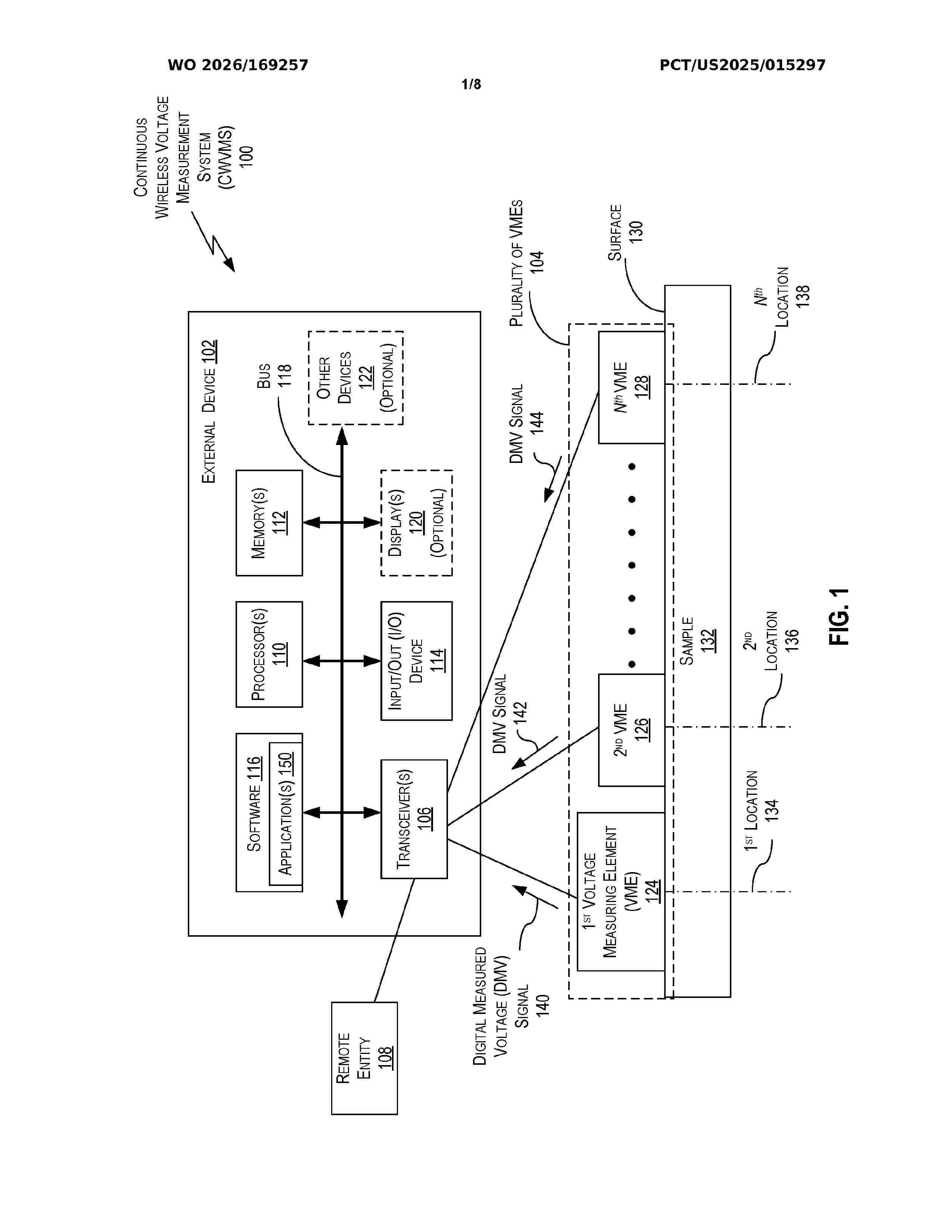
Techniques are discussed for a continuous wireless voltage measurement system (CWVMS). The CWVMS may comprise an external device having at least a first transceiver and a plurality of voltage measuring elements. Each voltage measuring element (VME) of the plurality of voltage measuring elements may include a voltage analysis element (VAE) configured to receive a measured voltage signal from a probe attached to a surface of a sample and convert the measured voltage signal to a digital measured voltage signal, and a voltage measuring element (VME) transceiver configured to wirelessly communicate with the external device and transmit the digital measured voltage signal to the external device; and each VME may be configured to produce a timestamp on the digital measured voltage signal.
Resumen de: WO2026169906A1
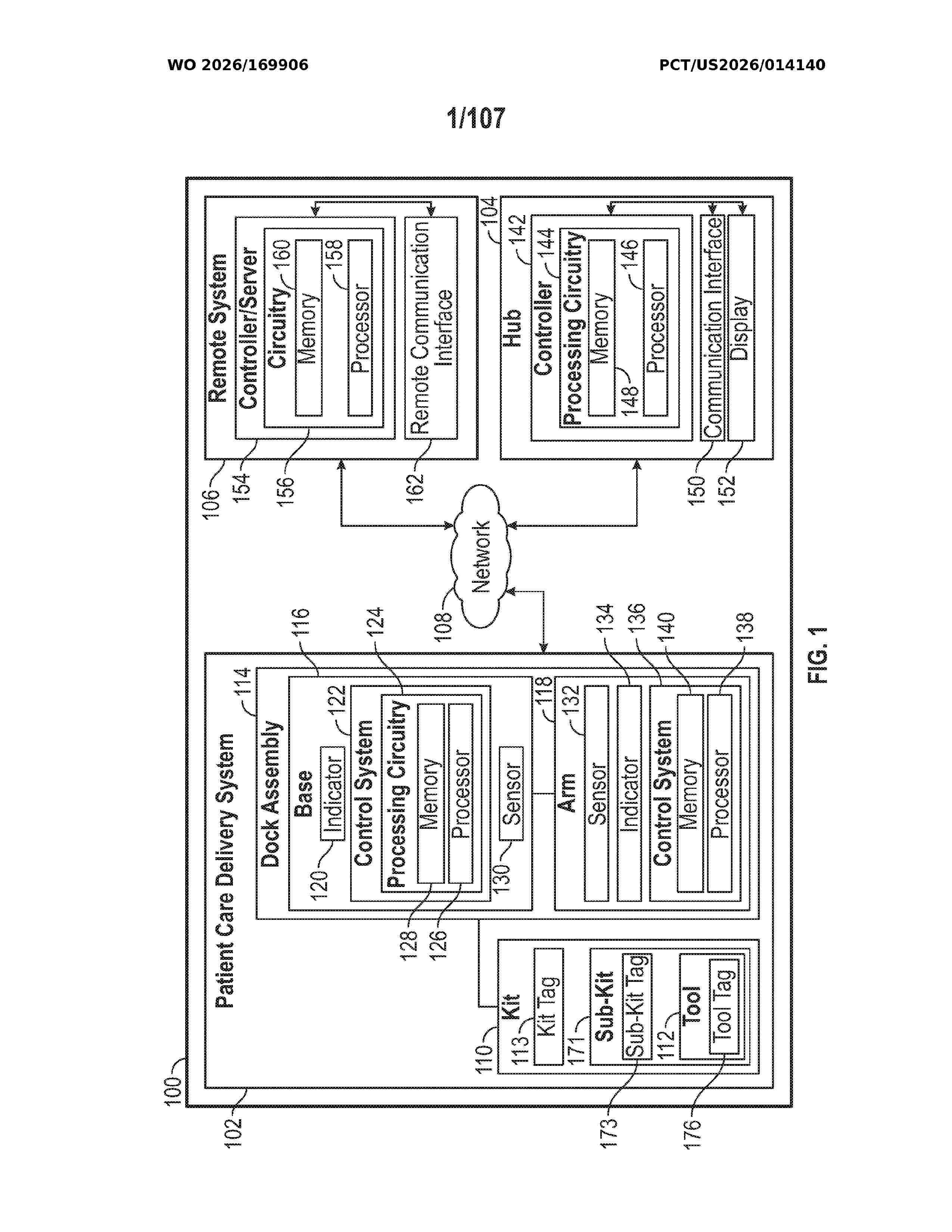
A patient care delivery system including a first kit including a first tool, and a dock assembly including a base, an arm coupled to the base and extending from the base, a first sensor disposed within the base or the arm, an indicator positioned on the base or the arm, and a processing circuit disposed within the base or the arm. The processing circuit includes a memory and one or more processors. The one or more processors are configured to receive a first signal from the first sensor when the first kit is at least one of coupled to the arm, suspended in front of the arm, or adjacent to the arm, after receiving the first signal, cause the indicator to provide a first indication to a user, and after not receiving the first signal, cause the indicator to provide a second indication to the user.
Resumen de: US20260235903A1
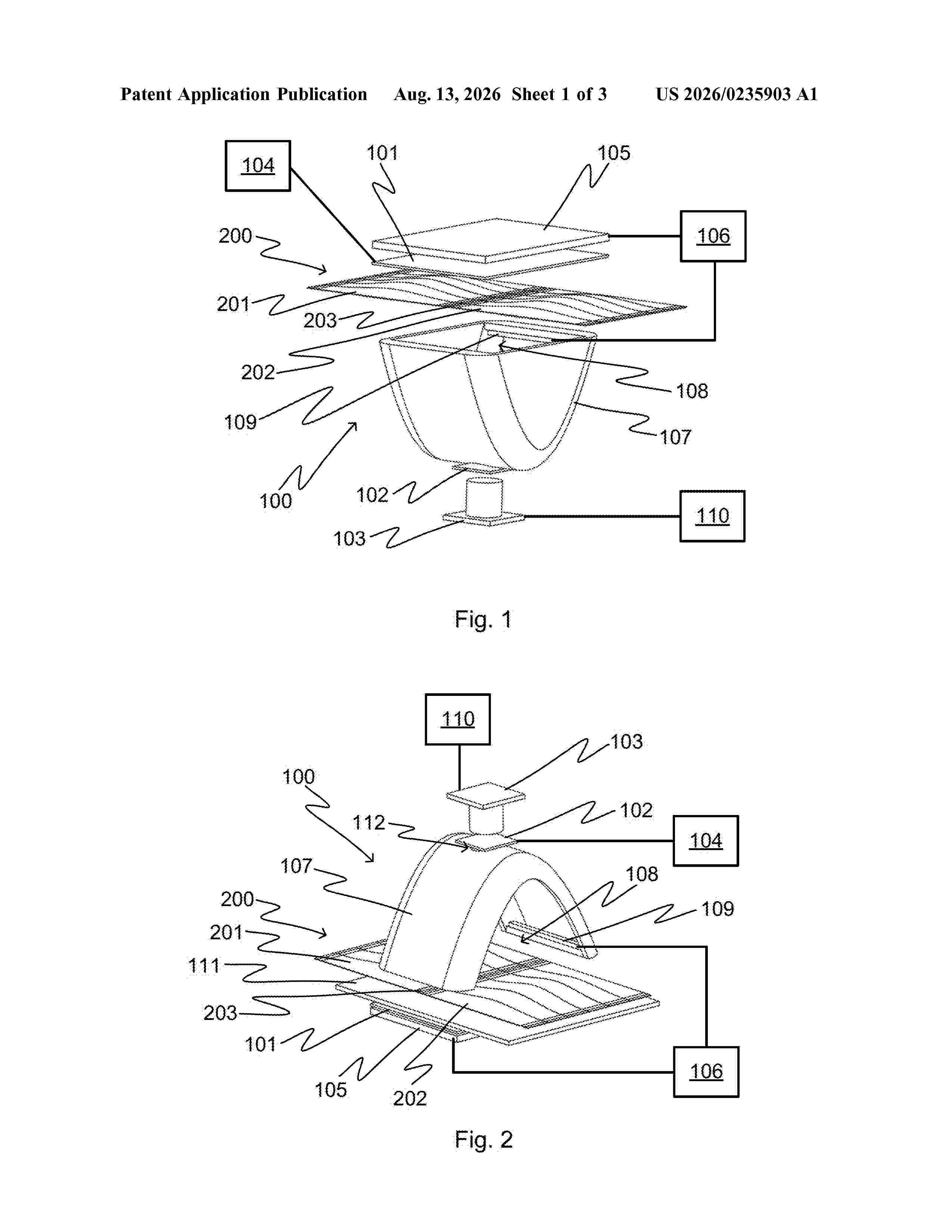
0000 An imaging device includes a first polarizer and a second polarizer between which a strip of medication sachets can be arranged. A camera is for imaging a first side of the strip of medication sachets through the second polarizer and a device for changing the polarization direction of the first polarizer or the second polarizer. A method is for detecting a seam area between medication sachets and to a medication dispenser.
Resumen de: US20260232191A1
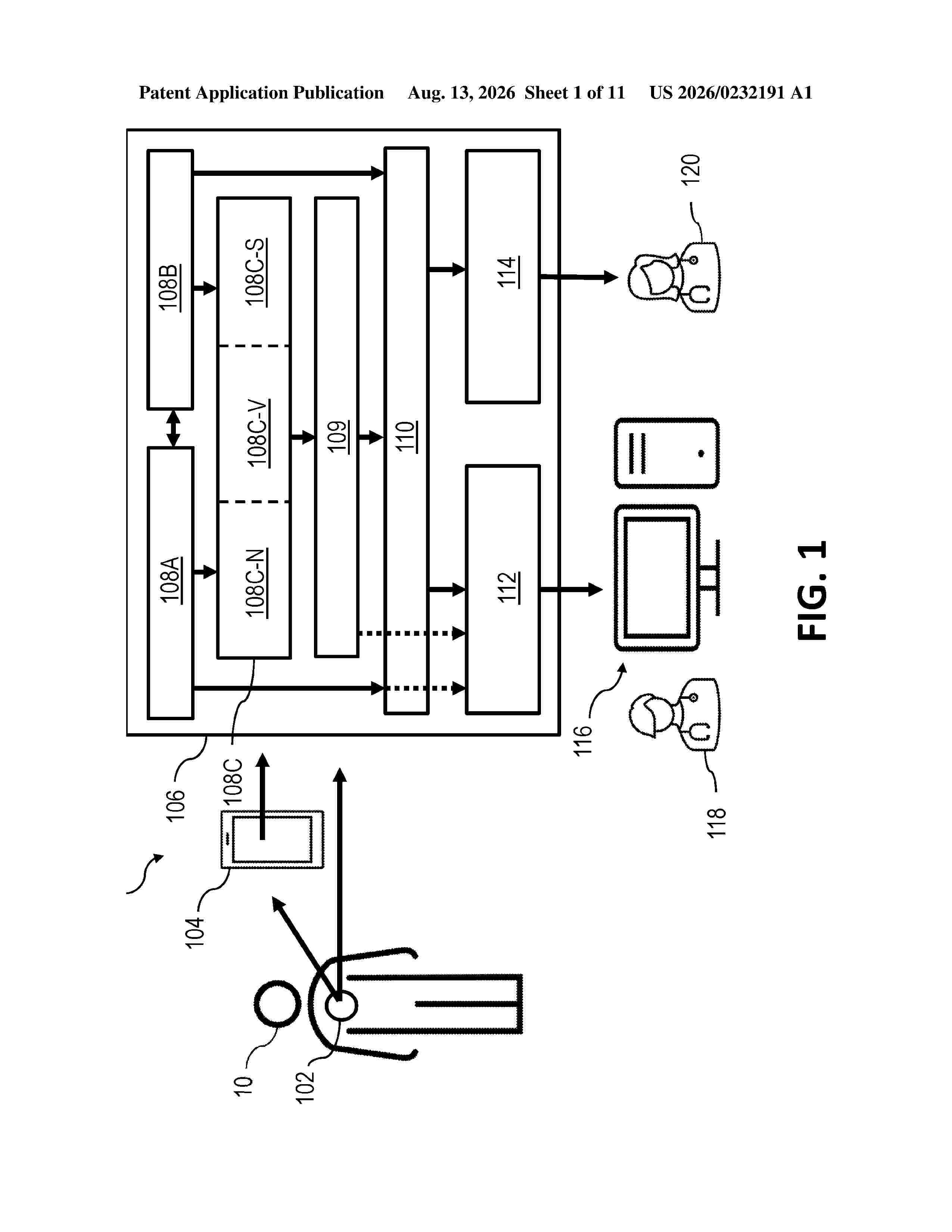
0000 Systems, methods, and devices involve approaches for tracking and displaying changes made to cardiac data. Certain approaches include altering metadata associated with time series cardiac data, generating an updated table to reflect the alterations, and displaying a change indicator in a first window on a user interface based on the changes to the metadata.
Resumen de: US20260235902A1
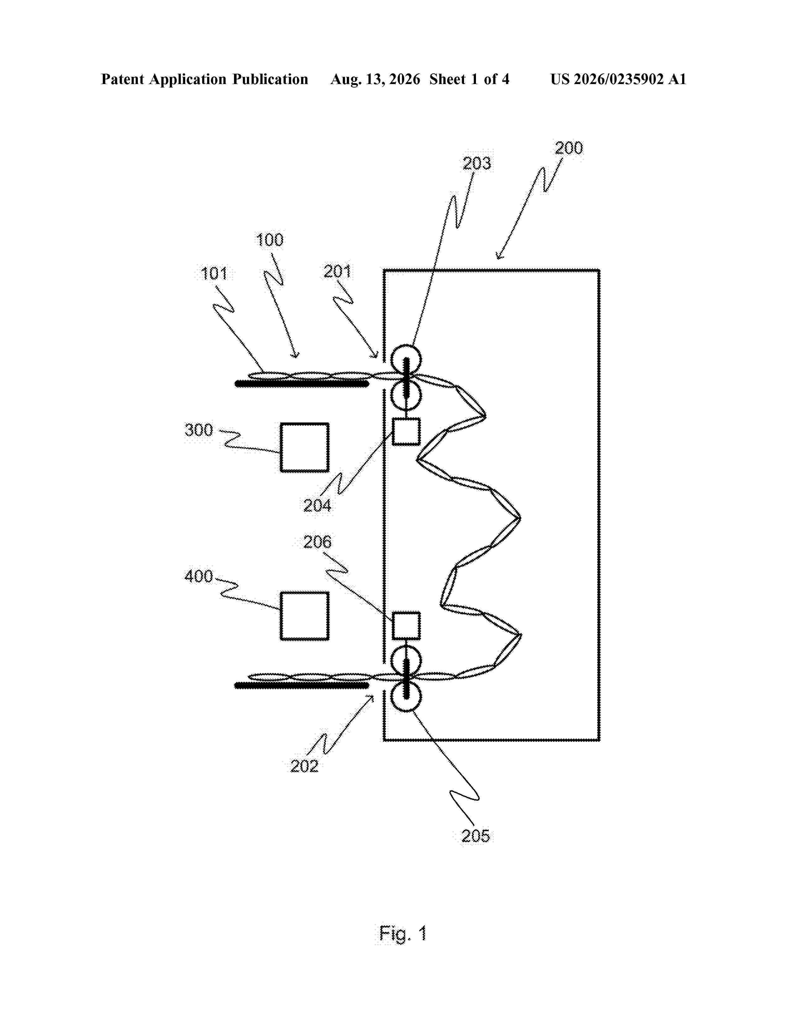
An arrangement is for handling a strip of medication sachets. The arrangement includes a conveying device for conveying the strip of medication sachets. The conveying device includes an inlet for receiving the medication sachets, an outlet for discharging the medication sachets and a conveyor for conveying the medication sachets from the inlet to the outlet. Aa first imaging device is arranged in connection with the inlet and configured to capture at least one image of the medication sachet before the medication sachet is conveyed to the inlet, and a second imaging device is arranged in connection with the outlet and configured to capture at least one image of the medication sachet after the medication sachet has been conveyed out of the outlet. A dispenser is for dispensing the medication.
Resumen de: WO2026169900A1
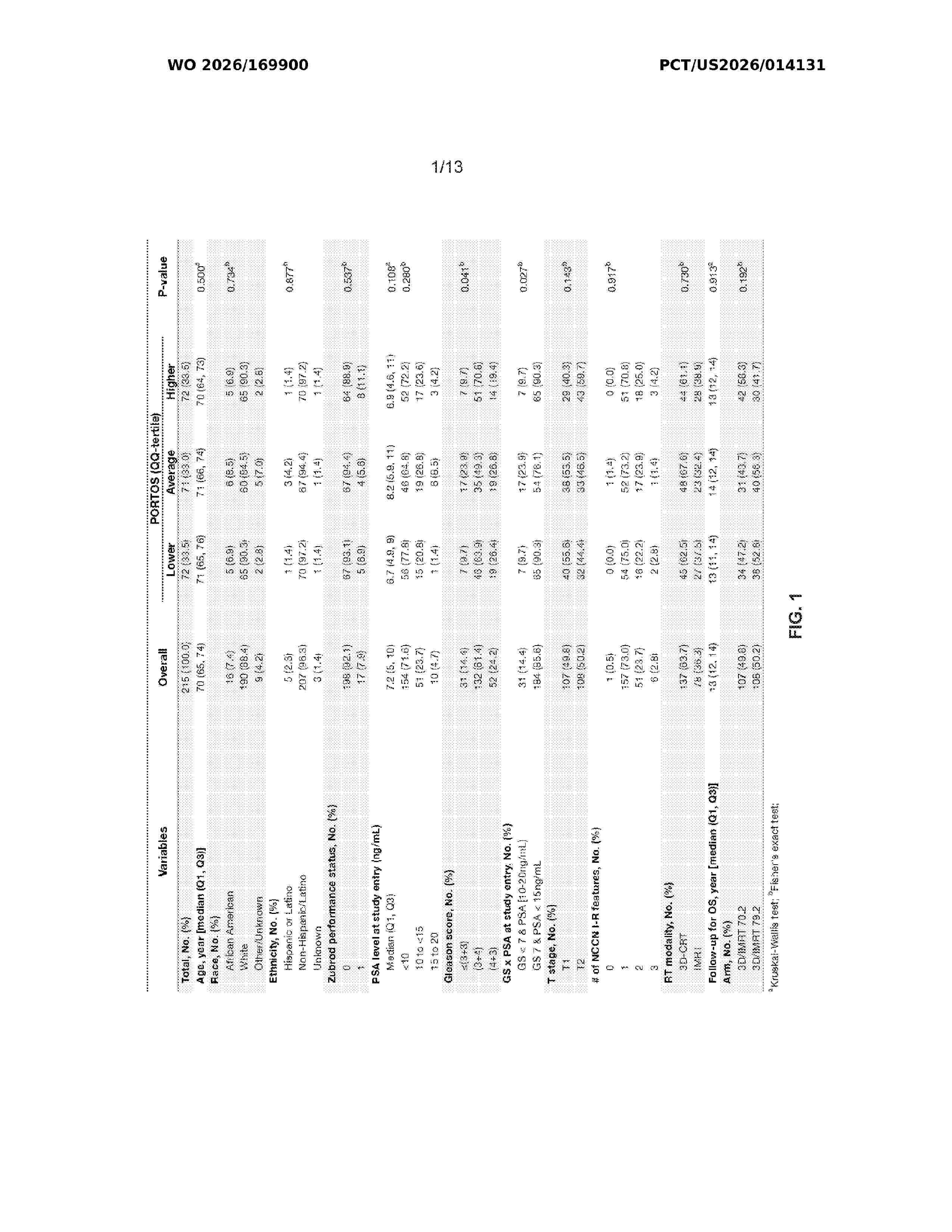
Provided herein are biomarkers for calculating a risk score (e.g., a PORTOS score) and/or determining an expected response to treatment comprising dose-escalated radiation therapy in a subject with prostate cancer who has not undergone radical prostatectomy (e.g., intermediate risk prostate cancer), methods of using the biomarkers including methods of treatment based on the risk score and/or expected response to treatment comprising dose-escalated radiation therapy determined using the biomarkers, as well as kits for detecting the biomarkers.
Resumen de: US20260232541A1
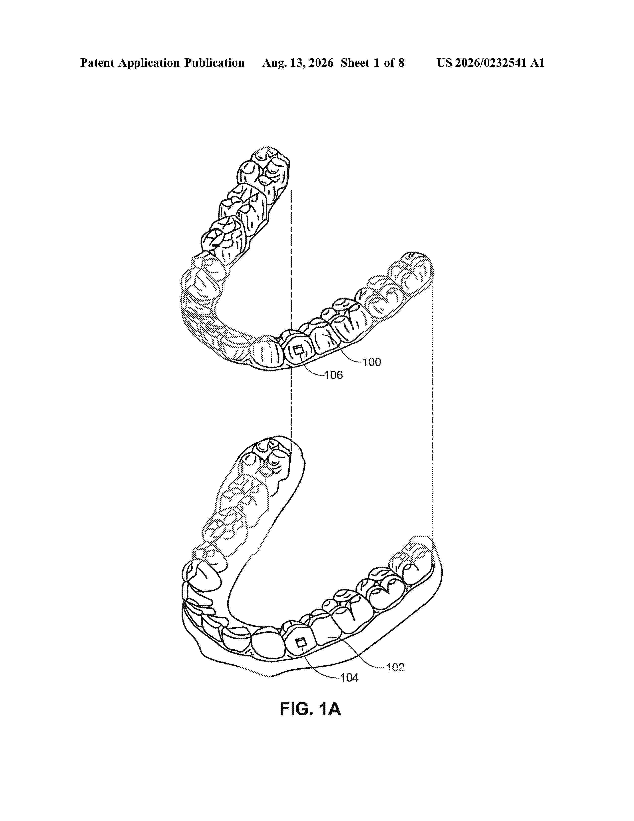
The present disclosure provides novel oligomers and polymerizable compositions comprising the same. Further provided herein are methods of producing polymers, polymer coatings, and devices. Also provided herein are methods of using polymer materials and polymer materials fabricated via additive manufacturing processes (e.g., via 3D printing), such as orthodontic appliances.
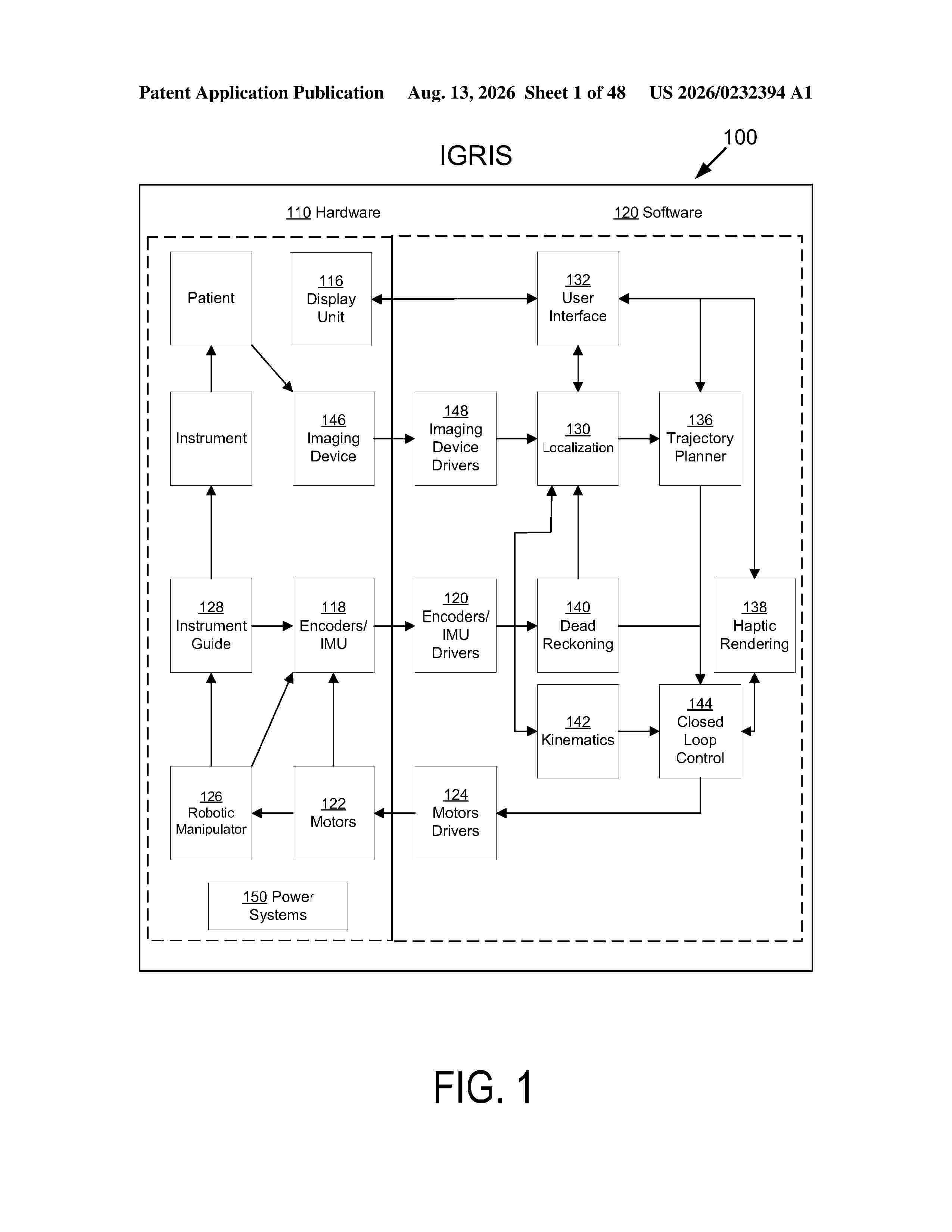
Resumen de: US20260232394A1
0000 An image-guided robotic intervention system ("IGRIS") may be used to perform medical procedures on patients. IGRIS provides a real-time view of patient anatomy, as well as an intended target or targets for the procedures, software that allows a user to plan an approach or trajectory path using either the image or the robotic device, software that allows a user to convert a series of 2D images into a 3D volume, and localizes the 3D volume with respect to real-time images during the procedure. IGRIS may include sensors to estimate pose of the imaging device relative to the patient to improve the performance of that software with respect to runtime, robustness, and accuracy.
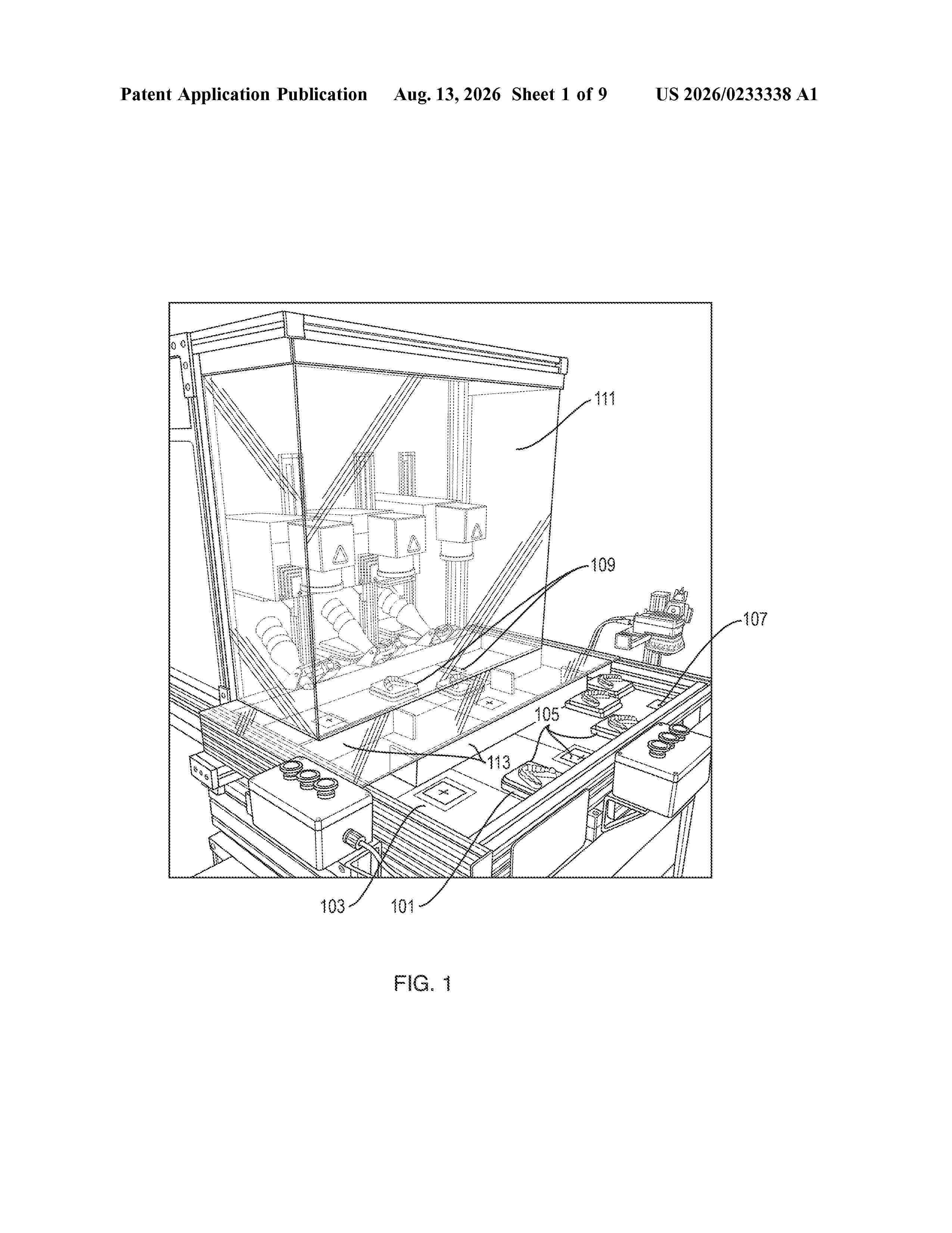
Resumen de: US20260233338A1
The present disclosure discusses techniques for marking a dental appliance. A dental model and dental appliance are positioned onto a conveyor tray, which is transported to a scan station. The dental model and conveyor tray each include an identifying code. The scan station includes a camera and barcode reader to read the identifying codes on the dental model and conveyor tray, and these codes are associated with one another. The conveyor tray then moves to a marking station where a barcode reader reads the code on the conveyor tray, and then marks the dental appliance with the appropriate marking.
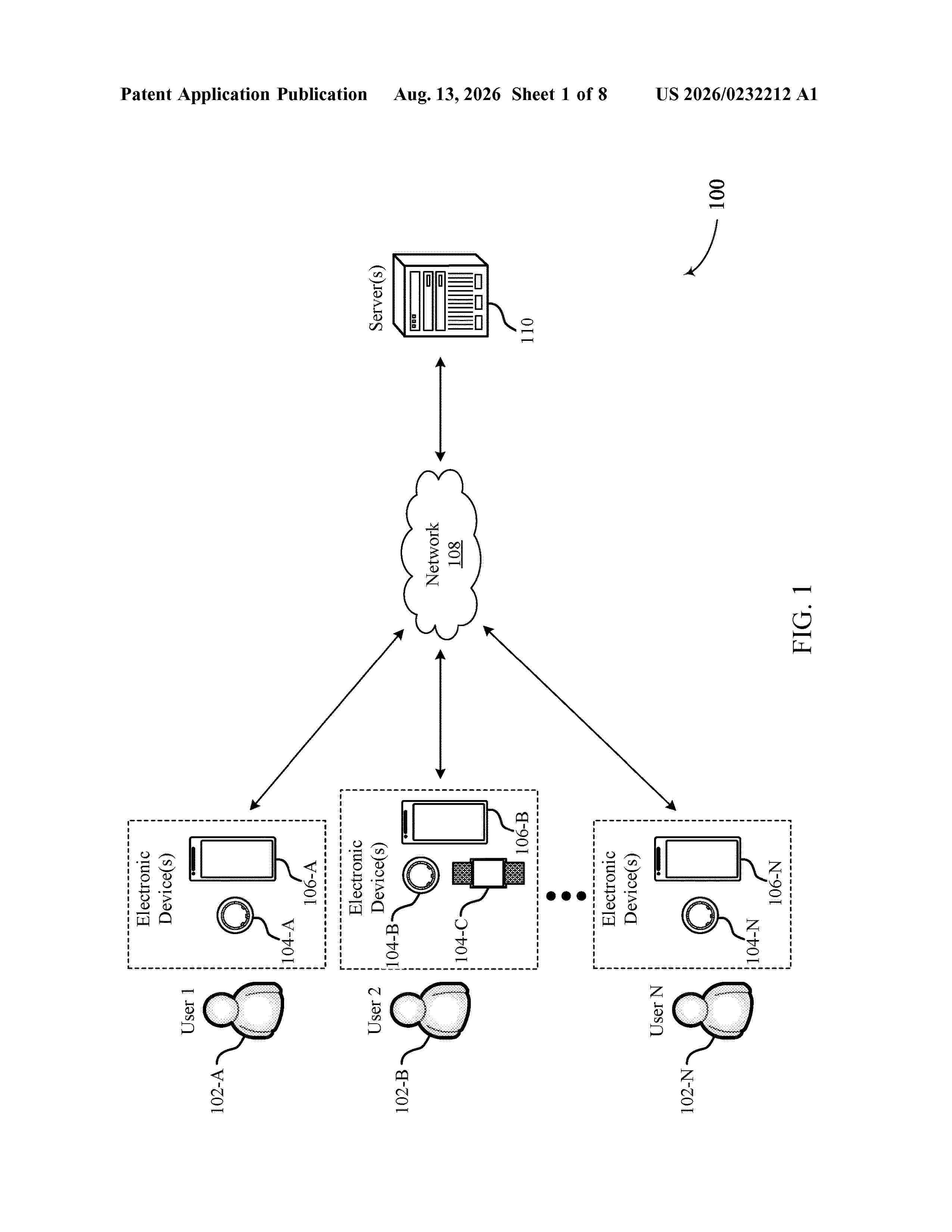
Resumen de: US20260232212A1
0000 Methods, systems, and devices for identifying representative photoplethysmogram (PPG) pulses are described. A method may include acquiring a first set of PPG pulses from a user via a wearable device. The method may include comparing a set of morphological features of the first set of PPG pulses, and determining one or more PPG profiles for the user, where the one or more PPG profiles each include a set of morphological value ranges for the set of morphological features. The method may include acquiring a second set of PPG pulses from the user via the wearable device, and determining that one or more PPG pulses from the second set of PPG pulses match the one or more PPG profiles. The method may include determining one or more physiological metrics associated with the user based on the one or more PPG pulses matching the one or more PPG profiles.
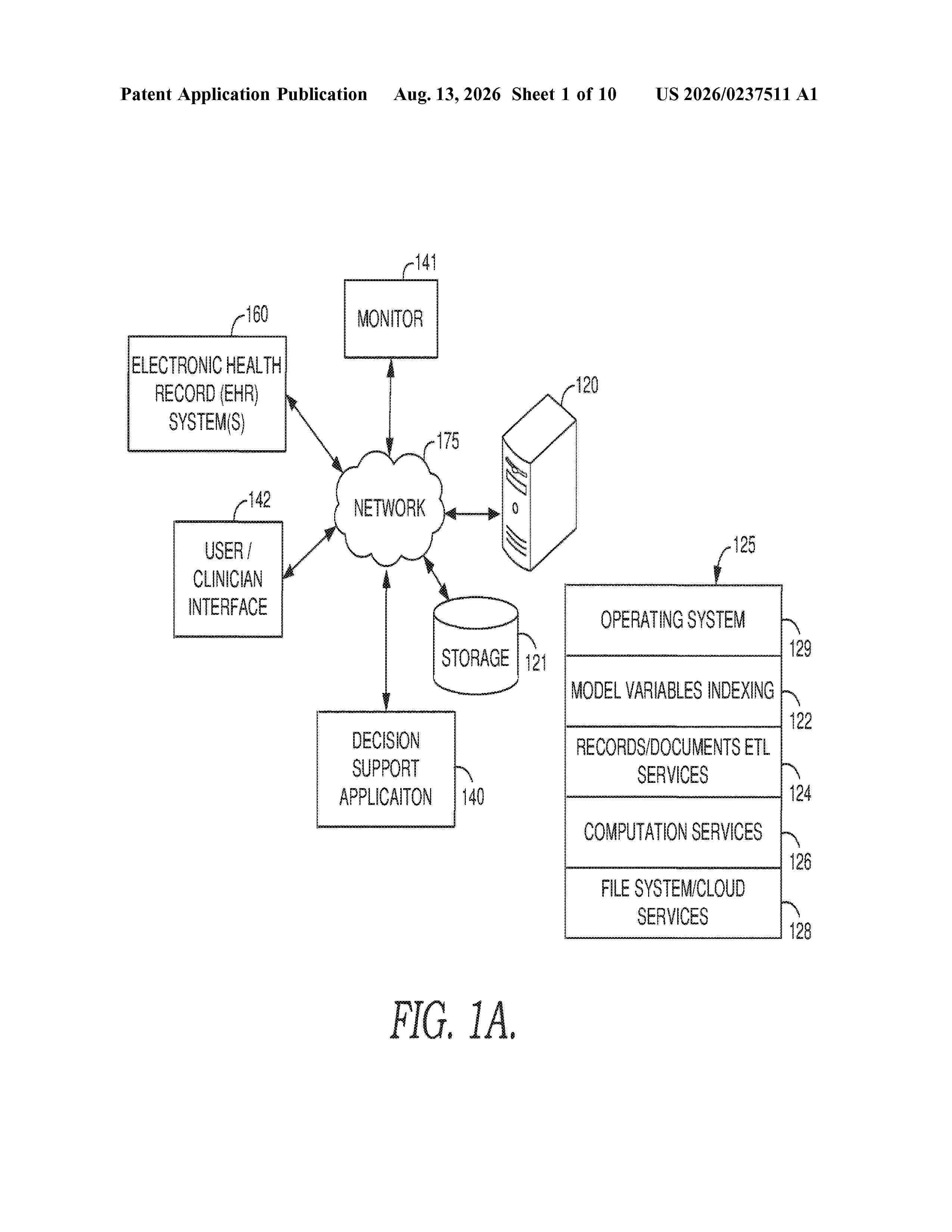
Resumen de: US20260237511A1
A decision support tool is provided for identifying and assisting clinicians with patient ventilator asynchrony. The information used to make the identification may include data from a patient's ventilator including the volume, flow, and pressure associated with that ventilator. At least some of this information may be used to compute one or more features for a time series of the data received for the patient. These features may be used in connection with heuristic rules and machine learning algorithms to identify instances of patient ventilator asynchrony. Based on the identification, one or more intervening actions may be initiated to reduce the impact of patient ventilator asynchrony.
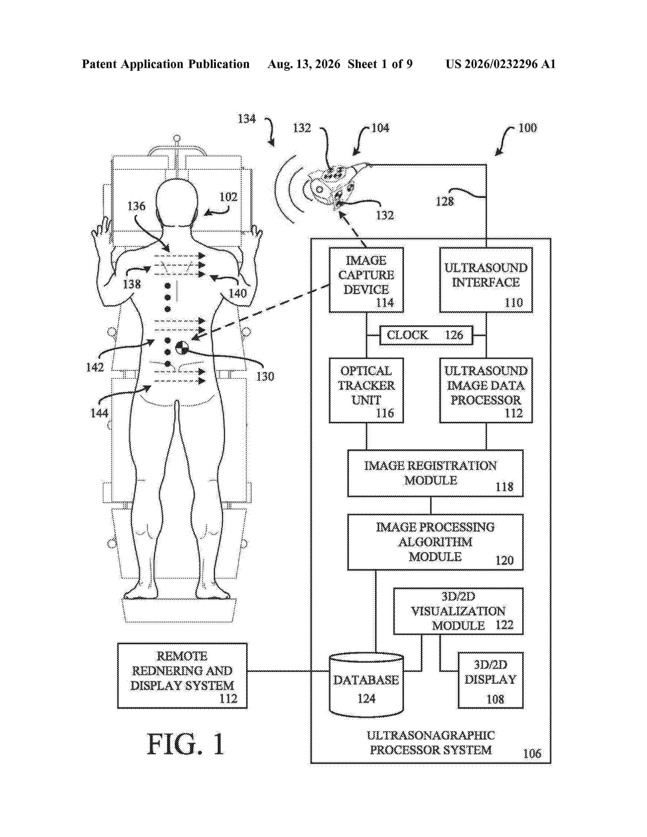
Nº publicación: US20260232296A1 13/08/2026
Solicitante:
VERDURE IMAGING INC [US]
Verdure Imaging, Inc.
Resumen de: US20260232296A1
A system and method for performing ultrasound scans is provided. One embodiment of the ultrasonographic system acquires sonogram information from a series of ultrasonic scans of a human subject. The series of ultrasound scans are taken over a portion of interest on the human subject which has their underlying bone structure or other ultrasound discernable organ that is under examination. The data from the series of scans are synthesized into a single data file that corresponds to a three-dimensional (3D) image and/or 3D model of the underlying bone structure or organ of the examined human subject.
 BOPI
BOPI
 Sede Electrónica