

Si deseas distinguir tus productos, servicios o ambos de los de otra empresa, es posible que necesites una marca o nombre comercial. Descubre qué son, en qué consiste su procedimiento de registro y qué implica.

Información sobre los plazos de presentación de solicitudes de transformación de marcas de la Unión Europea en marca nacional española. Más información


Si tienes un nuevo dispositivo, producto o procedimiento que resuelva un problema técnico o tenga una ventaja práctica, existen distintas formas de protegerlo en España y en otros países. Descubre cómo hacerlo.

¿Tu innovación reside en la estética, la ornamentación o la apariencia de tu producto? Protégela mediante un diseño industrial. Descubre qué derechos confiere el registro y cómo realizar la tramitación.

Las indicaciones geográficas protegen el nombre de un producto originario de una zona geográfica, a la cual le debe una determinada calidad, reputación u otra característica. Descubre qué son, en qué consiste su procedimiento de registro y qué beneficios conceden.

Las patentes publicadas en todo el mundo son una valiosa fuente de información científica, técnica y comercial.

Si eres emprendedor/a o una empresa y quieres potenciar y mejorar la rentabilidad de tu negocio protegiendo de forma adecuada los activos intangibles de tu organización, en este espacio encontrarás lo necesario.


 169
resultados
169
resultados
 Última actualización
01/04/2026 [08:17:00]
Última actualización
01/04/2026 [08:17:00]


 Resultados 150 a 169 de 169
Resumen de: US20260060688A1
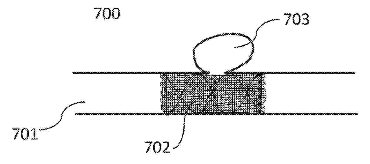
A biodegradable flow diverting device (BFDD) that will regulate blood flow into an aneurysmal sac, act as a scaffold for endothelization at the neck of an aneurysm, and degrade after successful dissolution of aneurysm and remodeling of blood vessel. This BFDD and associated fabrication method have the following features: (1) This is a non-braided FDD. The pore shapes, sizes, architectures (especially at the inlet and outlet of the pores), pore densities and porosities can be controlled for the optimum performance depending on the blood vessel and aneurysmal morphologies from patient MRI images, (2) BFDD is developed on a rotary arm with programmable variable speed and diameter in conjunction with a micromotion stage (3) Fabrication system can take any material including blended/composite biomaterials by adjusting temperature of the electro-melt extruder/needle and (4) Fabrication system is compatible with CAM (computer aided manufacturing) software and able to operate based on the adapted G-code.

Resumen de: US20260060750A1
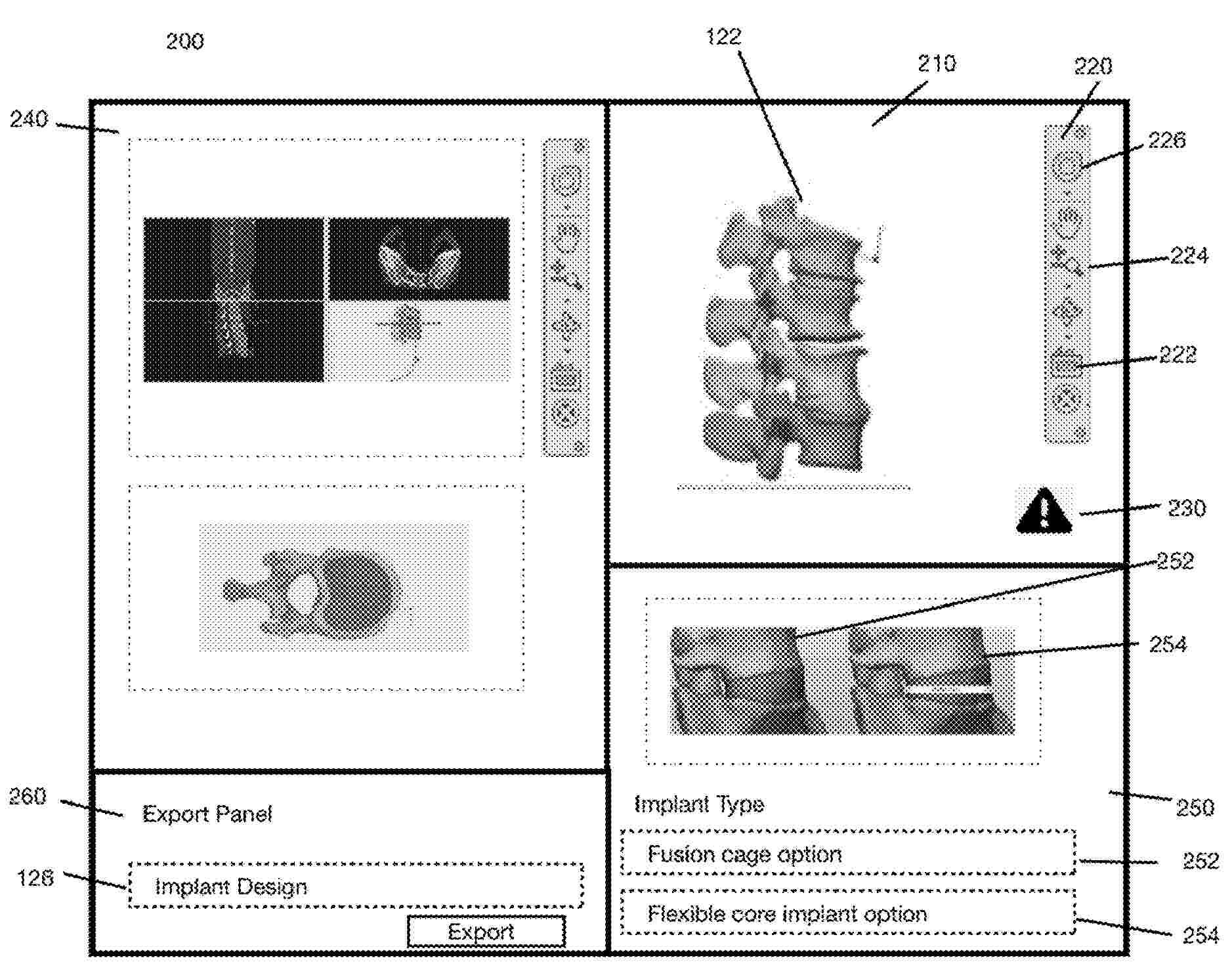
A system and method for planning and simulating a surgical operation to create a patient-specific spinal implant are disclosed. The system comprises a remote server configured to receive patient-specific medical image data and generate a 3D mesh model of the patient's spine using algorithms that separate vertebral bodies, remove artifacts, and smooth surfaces. A doctor's computer receives the 3D mesh model and allows real-time manipulation of intervertebral spaces to achieve a desired spinal curvature. The server generates a spinal implant design with surface-mapped endplates matching the patient's vertebral anatomy, which is transmitted to a 3D printer for manufacturing. The method includes steps of receiving image data, generating and updating the 3D mesh model based on doctor input, generating the final implant design, and transmitting it for production. The invention enables the creation of patient-specific spinal implants with improved conformity and surgical outcomes.
Resumen de: DE102024125325A1
1. Die Erfindung betrifft eine Membran (1) für einen Membrankontaktor (2), die als eine zu einer Mittelebene (ME) spiegelsymmetrische hohle Röhrenstruktur aus verzweigten Röhren (3, 3', 3", 3''', 3'''') mit beidseits der Mittelebene (ME) mehreren zur Mittelebene (ME) parallelen Verzweigungsebenen (VE) ausgebildet ist, wobei die Wandungen der Röhrenstruktur stoffpermeabel und/oder energiepermeabel sind und wobei im Bereich (B) jeder Verzweigungsebene (VE) jede in Richtung zur Mittelebene (ME) erstreckte Röhre (3, 3', 3'', 3''') in mehrere zur Mittelebene (ME) erstreckte Röhren (3', 3'', 3''', 3'''') verzweigt und die von beiden Seite der Mittelebene (ME) von der jeweils letzten Verzweigungsebene (VE) zur Mittelebene (ME) erstreckten Röhren (3'''') im Bereich (ÜB) der Mittelebene (ME) ineinander übergehen. Die Erfindung betrifft weiterhin einen Membrankontaktor (2) mit einer solchen Membran und ein Verfahren zur Herstellung einer hohlen Membran (1).
Resumen de: US20260060845A1
A wearable wound management system integrating a flexible microfluidic assembly and real-time electrochemical sensing to autonomously sample, transport, and analyze wound exudate. A Janus membrane, formed by selective deposition of perfluoroalkyl-functionalized silica nanoparticles and O2 plasma etching on a PET film, may collect fluid via its superhydrophobic wound-facing side and deliver it to a curved, wedge-shaped microfluidic channel that enhances capillary flow. Downstream, a graded PDMS micropillar array refreshes a sensing region by unidirectional fluid movement. A drop-on-demand inkjet-printed, CO2 laser-patterned flexible sensor patch may measure nitric oxide, oxygen, hydrogen peroxide, pH, temperature, and other relevant metrics. An encapsulated wireless electronic module may transmit health data for wireless monitoring. This system, combined with machine-learning analytics, may enable continuous, in situ monitoring and predictive wound classification, supporting proactive and personalized chronic wound care.
Resumen de: WO2026049910A1
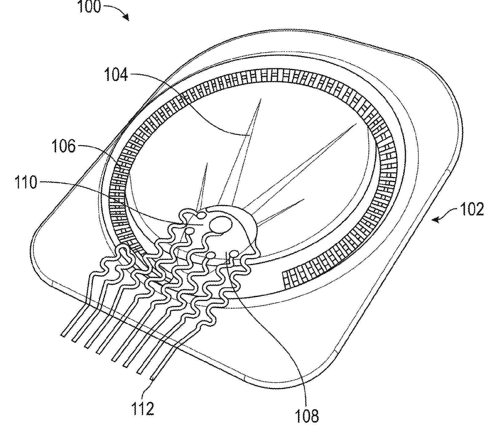
Embodiments relate to bioelectrodes designed to accommodate patient specific gyral patterns, and to methods of making and using thereof. A bioelectrode may be manufactured by obtaining a three-dimensional scan of at least a portion of a brain; identifying a target region of the brain; designing a bioelectrode model configured to fit the target region; and printing the bioelectrode based on the bioelectrode model. The bioelectrode is printed to include a top layer, a bottom layer, and an electrode layer positioned between the top layer and the bottom layer such that the top layer and bottom layer encapsulate the electrode layer. The bioelectrode may have a Young's modulus between 0.1 and 10 kPa to better mechanically match brain tissue.
Resumen de: US20260060783A1
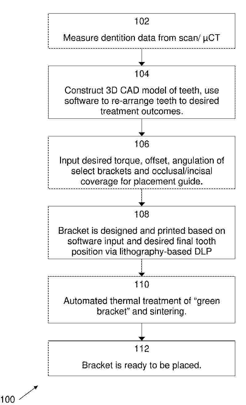
In an embodiment, a method of manufacturing customized ceramic labial/lingual orthodontic brackets by additive manufacturing may comprise measuring dentition data of a profile of teeth of a patient, based on the dentition data, creating a three dimensional computer-assisted design (3D CAD) model of the patient's teeth, and saving the 3D CAD model, designing a virtual 3D CAD bracket structure model for a single labial or lingual bracket structure based upon said 3D CAD model, importing data related to the 3D CAD bracket structure model into an additive manufacturing machine, and directly producing the bracket with the additive manufacturing machine by layer manufacturing from an inorganic material including at least one of a ceramic, a polymer-derived ceramic, and a polymer-derived metal.
Resumen de: KR20260029152A
본 발명 합성수지 라미네이트 제조를 통한 예측가능하고 정확한 라미네이트 기공물 제조방법은 치아 일부를 삭제한 후 세라믹 소재의 기공물을 삭제된 치아에 부착하는 방식으로 이루어지는 치과시술 시에 피시술자의 만족도를 향상시키고 빠르고 정확하게 라미네이트 치과시술을 실시할 수 있는 라미네이트 기공물 제작 방법에 관한 것으로, 피시술자의 삭제된 치아를 포함하는 구강 내의 3D 스캔 정보를 취득하는 구강 3D 스캔 데이터 획득 단계와, 치과용 3D 디자인 프로그램을 통하여 다수의 기공물을 디자인하여 다수의 기공물 출력데이터를 생성하는 다수의 기공물 출력데이터 생성 단계와, 합성수지 소재 출력 치과용 3D 프린터를 이용하여 다수의 합성수지 기공물을 출력하는 단계와, 합성수지 기공물 핏팅 과정을 통해 선택된 어느 하나의 합성수지 기공물의 기공물 출력데이터를 세라믹 기공물 출력데이터로 결정하는 세라믹 기공물 출력데이터 결정 단계와, 세라믹 소재 출력 치과용 3D 프린터를 이용하여 세라믹 소재의 기공물을 제작하는 세라믹 기공물 제작 단계를 포함하는 것을 특징으로 하여, 다수의 기공물 핏팅 과정을 통해 선택된 디자인의 세라믹 소재의 기공물을 제작함에 따라 피시술자의 불만족에 따른 디�
Resumen de: KR20260029070A
본 발명은 3D 프린팅용 임시치관 레진에 관한 것으로서, 보다 구체적으로는 환자의 치아 색상과 같이 자연스러운 색상을 가지며 내구성이 우수한 임시치관을 정밀하고 빠르게 제작할 수 있는 3D 프린팅용 임시치관 레진 및 그 임시치관 레진으로 제조한 임시치관의 제조방법에 관한 것이다.
Resumen de: WO2024224174A1
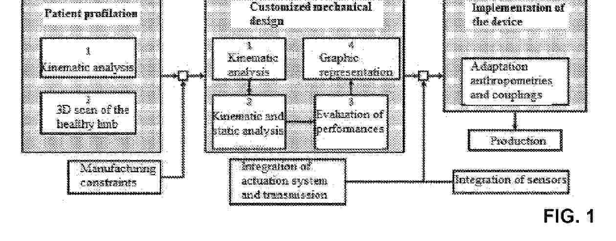
A method for designing a functional prosthesis of customized hand, for patients amputated at trans-radial level, implemented with additive manufacturing technology. The method flow for designing and developing the devices consists of the following steps: i) kinematic analysis and 3D scan of the limb contralateral to the amputated one; ii) kinematic synthesis of the planar mechanism at one active degree of freedom (GdL), modular for each finger, and optimization of the same through a global performance index (GPI); iii) prototyping of the assembly of five fingers.
Resumen de: WO2024223794A1
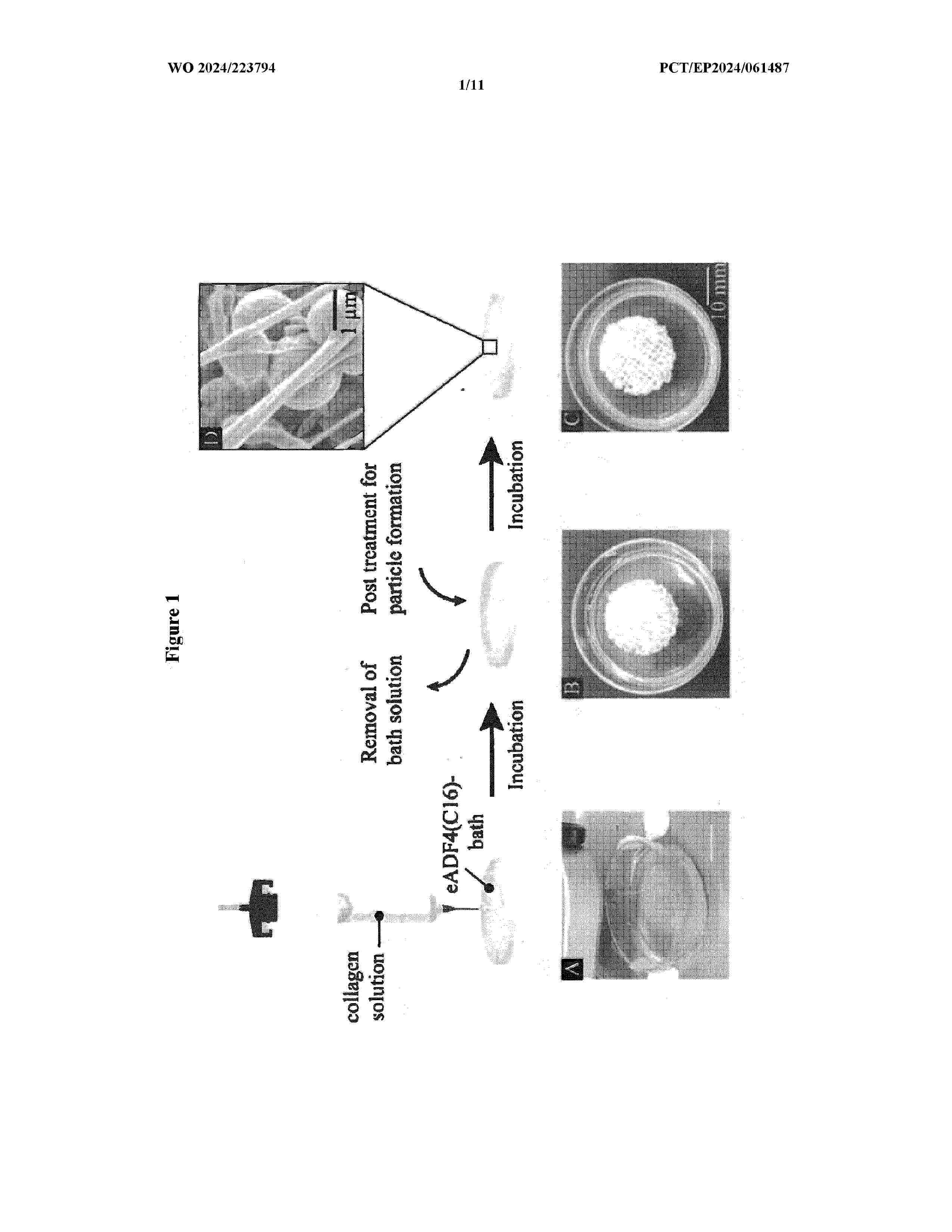
The present invention relates to a method for producing a collagen and silk polypeptide composite structure. In addition, the present invention relates to a collagen and silk polypeptide composite structure and uses thereof.
Resumen de: WO2024226028A1
A band wrapping device forms a length of band material into a loop for automatically applying the band to a limb (e.g., wrist) of a user. The band wrapping device may receive the length of band material from a cassette receivable in the band wrapping device and can include a band transport mechanism for transporting the band material, a sealer module for sealing the band material to itself and cutting the band material to a length, and/or a ring module for reception of the limb of the user for band application. The device and its constituent components can help reduce the contact between the individual requiring a band and those applying the band, can help reduce waste by using a more precise amount of band material sized to the limb, and can help reduce the staffing requirements in areas in which the bands are to be applied.
Resumen de: CN121588283A
本发明公开了一种基于甲基丙烯酰化明胶的微球、其制备方法及其在生物墨水中的应用,该微球以甲基丙烯酰化明胶为核心基材,可选择性复合糖胺聚糖‑甲基丙烯酰化明胶和/或脂质体,通过微流控技术制备而成,所得微球尺寸均一,且复合组分可赋予微球优异的自粘附特性;进一步采用微流控技术,以基材及复合组分作为微球壳相、细胞分散液作为微球核相,制备具有核壳结构的载细胞微球,该载细胞微球能良好维持包埋细胞的正常存活与生长。基于甲基丙烯酰化明胶的微球或载细胞的基于甲基丙烯酰化明胶的微球,可作为3D打印墨水用于制备支架材料、载细胞支架材料,适配细胞三维培养、细胞球培养、高密度细胞培养及组织工程植入材料领域。
Resumen de: WO2025027496A1
A custom tool for forming a dental restoration in a mouth of a patient includes a one-piece mold body providing for a customized fit with at least one tooth of the patient, the one-piece mold body including an occlusal portion forming an occlusal surface corresponding with an occlusal surface of the tooth, a mesial proximal portion forming a mesial proximal surface corresponding with a mesial proximal surface of the tooth, and a distal proximal portion forming a distal proximal surface corresponding with a distal proximal surface of the tooth. The mold body is configured to combine with the tooth of the patient to form a mold cavity encompassing missing tooth structure of the tooth. The occlusal portion, the mesial proximal portion, and the distal proximal portion are based on three-dimensional scan data of the mouth of the patient.
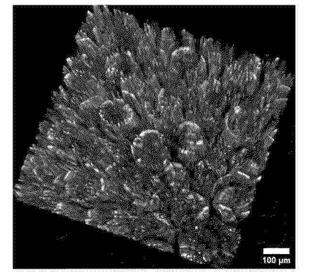
Resumen de: CN121592153A
本发明公开了一种3D打印用的医用级PEEK复合材料及其制备方法,涉及3D打印技术领域,解决了现有PEEK材料存在的生物活性低的问题。该医用级PEEK复合材料由以下重量份的原料制成:聚醚醚酮基体80~95份、生物活性填料5~15份、界面改性剂0.5~5份、导热与成核增强剂0.1~3份;所述生物活性填料包括重量比为(1~5):(5~1)的纳米羟基磷灰石和生物活性玻璃。能够有效模拟天然骨环境,提高复合材料的生物活性,促进细胞黏附与增值,从而提高细胞增值率,还能有效提高了复合材料的抑菌效果和力学性能。
Resumen de: CN121587868A
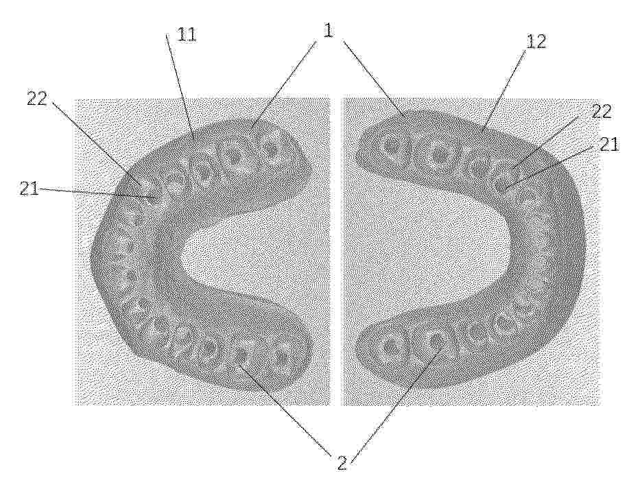
本发明公开了临时修复体穿龈三维形态塑形工具盒及其制造方法。包括本体和塑形凹槽,塑形凹槽开设于所述本体上,塑形凹槽用于提供种植替代体的放置空间并模拟天然牙穿龈三维形态的结构;其中,本体包括上本体和下本体;上本体和下本体可拆卸连接;塑形凹槽包括种植替代体放置结构和穿龈轮廓成型结构,种植替代体放置结构与穿龈轮廓成型结构周边连接,种植替代体放置结构用于放置与种植体匹配的种植替代体,穿龈轮廓成型结构用于容纳与种植替代体相连接的临时修复基台,并模拟天然牙穿龈三维形态。
Resumen de: WO2025027496A1
A custom tool for forming a dental restoration in a mouth of a patient includes a one-piece mold body providing for a customized fit with at least one tooth of the patient, the one-piece mold body including an occlusal portion forming an occlusal surface corresponding with an occlusal surface of the tooth, a mesial proximal portion forming a mesial proximal surface corresponding with a mesial proximal surface of the tooth, and a distal proximal portion forming a distal proximal surface corresponding with a distal proximal surface of the tooth. The mold body is configured to combine with the tooth of the patient to form a mold cavity encompassing missing tooth structure of the tooth. The occlusal portion, the mesial proximal portion, and the distal proximal portion are based on three-dimensional scan data of the mouth of the patient.
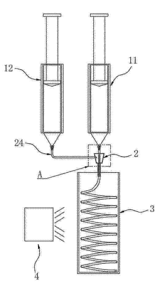
Resumen de: CN121590022A
本发明涉及人工血管制造的技术领域,具体而言,公开了一种人工血管打印方法及设备包括:第一储液部件,用于储存芯相材料;第二储液部件,用于储存外相材料;同轴打印组件,具备同轴设置的内料管和外料管,内料管连接第一储液部件,外料管连接第二储液部件;压力源,连通第一储液部件和第二储液部件,用于提供压力;接收部件,设置于同轴打印组件下方,用于接收人工血管;光固化组件,设置于接收部件的一侧,用于固化人工血管;通过使用本发明,能够复现天然管状组织复杂的解剖学特征,能够防止微小血栓的产生;且能够有效避免造成微通道堵塞,避免影响制备精度和效率,能够兼顾血管结构的生物相容性和高机械强度。
Resumen de: CN121592122A
本发明公开了一种用于挤出3D打印的光‑酸协同增强生物墨水及其制备方法。该墨水由N‑乙烯基吡咯烷酮(NVP)、聚乙烯醇(PVA)、光引发剂及具有“单羟基‑双羧基”特异结构的苹果酸组成。打印时,墨水经挤出并结合紫外光照射实现初步定型,随后在加热油性支撑浴中同步触发酸催化NVP深度聚合及PVA微晶化,一步完成光固化定型与热处理增强。本发明利用苹果酸独特的分子结构构建了高效的光‑酸协同增强机制,不仅显著提升了单体转化率,更通过物理‑化学双网络大幅增强了打印体的力学强度与形状保真度,有效解决了生物墨水可打印性与强度的矛盾,在组织工程、柔性电子等领域具有广阔应用前景。
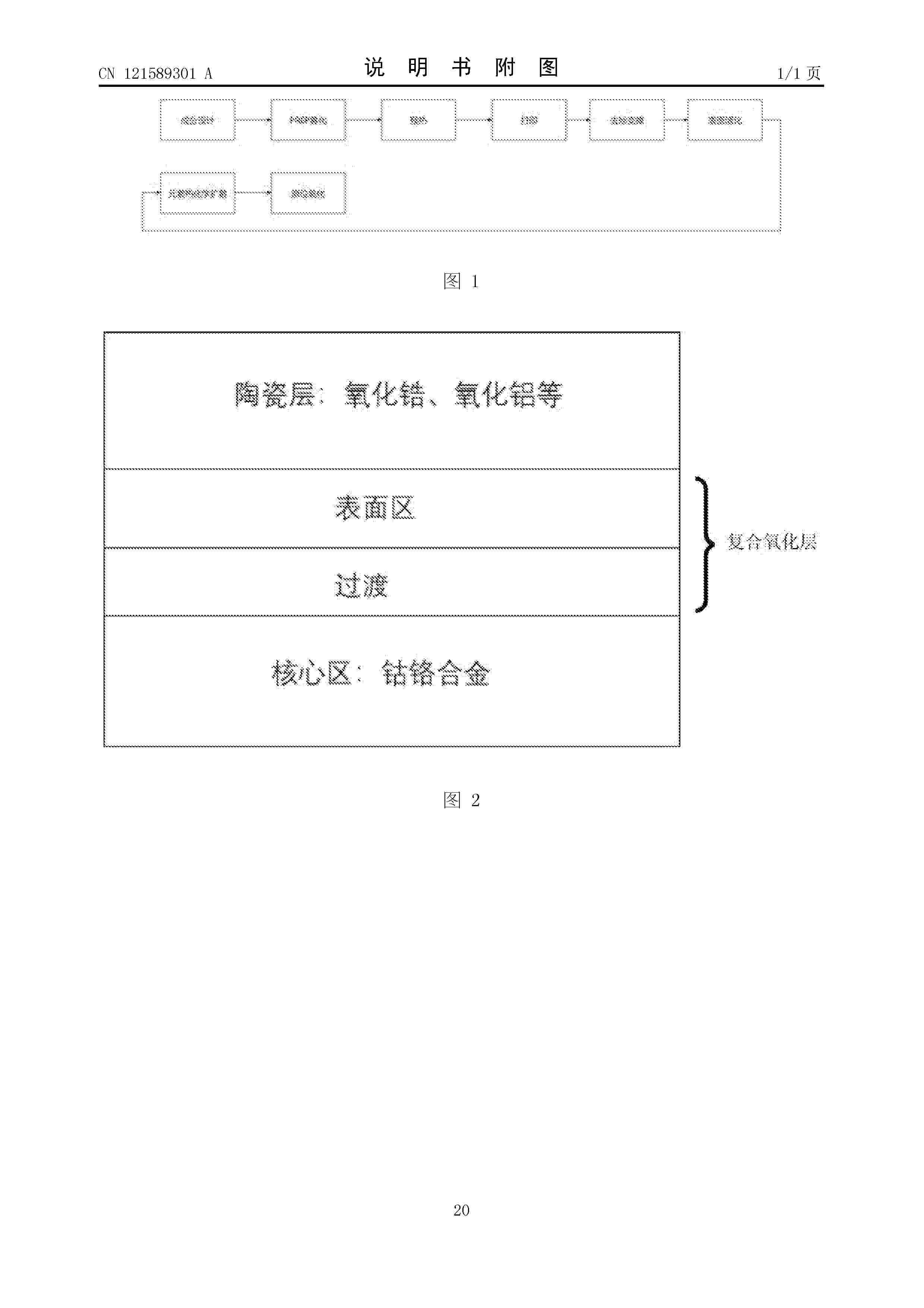
Nº publicación: CN121589301A 03/03/2026
Solicitante:
上海前研高合新材料科技有限公司
Resumen de: CN121589301A
本发明的目的在于提供一种降低裂瓷率的齿科用钴铬合金修复体3D打印工艺,其中,钴铬合金粉末按重量百分比包括如下成分:Co:58‑64wt%;Cr:25‑31 wt %;Mo:5‑6 wt %;W:2‑3 wt %;Si:0.8‑1.2 wt %;Mn:0.5‑1.0 wt %;稀土氧化物:0.1‑0.3 wt %。打印工艺包括:采用如本发明所述的钴铬合金粉末预热、进行3D打印,打印过程中通过动态能量密度调制分别打印核心区、过渡区和表面区后获得修复体打印件,将钴铬合金打印件进行后处理获得钴铬合金修复体。本发明突破性结合粉末材料,3D打印工艺和后处理元素掺杂技术,显著提高金瓷结合力、实现金属与陶瓷的渐进式连接,从本质上解决界面失配问题。
 BOPI
BOPI
 Sede Electrónica